| Service | 🇺🇦 Ukraine | 🇨🇿 Czech Republic | 🇹🇷 Turkey |
|---|---|---|---|
| Wisdom tooth removal | from USD 52 | from USD 143 | from USD 167 |
| CT scan, 2 jaws (for clinic patients) | from USD 15 | — | — |
| Computer-controlled anaesthesia | from USD 13 | from USD 7 | from USD 7 |
| Drug sedation, 30 min | from USD 33 | — | — |
Prices in the “Ukraine” column come from MediUkraine’s partner clinics. The Czech Republic and Turkey columns show the lower bounds of prices published by clinics there (for Turkey, also the Turkish Dental Association’s recommended 2026 tariff), collected in August 2026 and converted to US dollars at the rate on the day of collection. A dash means we could not find a comparable published price. In any country, the final cost is set by the clinic after an examination.
Impaction is the condition in which a tooth has fully formed but never erupted into the dental arch and has stayed in the bone or under the gum. If only part of the crown has come through, it is called partial impaction. A related term, malposition, describes a tooth in the wrong position: tilted forwards or backwards, rotated, displaced sideways or lying horizontally. Impaction and malposition often occur together, and the combination determines how complex the procedure will be.
The teeth most often left in the bone are the lower third molars, the wisdom teeth. Next in frequency come the upper canines: they fail to erupt in roughly 1–3% of people, and in most of these cases the tooth lies on the palatal side. Less often impacted are second premolars, lower canines and supernumerary teeth, the most common of which is the mesiodens, an extra tooth between the upper central incisors.
The indications for surgery are specific: recurrent pericoronitis over a partially impacted tooth; caries or root resorption of a neighbouring tooth caused by pressure from the unerupted crown; a follicular cyst around the tooth; a tooth that obstructs an orthodontic plan; preparation of the area for an implant or prosthetic work; a supernumerary tooth blocking the eruption of the incisors. A separate case is ankylosis, when the tooth fuses with the bone and can no longer be brought into the arch.
It is just as important to know when surgery is not needed. In an adult, a deeply impacted tooth with no symptoms, no cyst and no effect on its neighbours calls for monitoring, with an examination and an X-ray every year or two, rather than preventive surgery. An impacted upper canine in a teenager is usually brought into the arch with orthodontics rather than removed: this tooth carries chewing load and shapes the smile line, so keeping it is usually more important than removing it.
MediUkraine helps patients find and arrange treatment; it is not a clinic. Oral surgery appointments take place at the Vidnova dental clinic in Lviv, 10 Ivana-Teodoziia Kurovtsia Street. We agree the date, pass on the quote and support patients throughout, including those travelling from abroad. Whether surgery is needed is decided by the doctor after an examination and a three-dimensional scan.

Travel. Ukrainian airspace is closed, so there are no commercial flights. Patients reach Lviv overland, most often from Poland: Rzeszów and Kraków airports are the usual entry points, with a train, bus or private transfer onward to the border and on to the city. Lviv is roughly 70 km from the Polish border, the shortest crossing of any Ukrainian city.
Visas. Citizens of the EU, the United Kingdom and the United States do not need a visa for short stays in Ukraine. Rules depend on your citizenship, so check your own before booking.
Safety, plainly. Ukraine is at war. Air raid alerts occur in Lviv, and an appointment can be interrupted by one. Several governments advise against travel to Ukraine, and while that advice stands, ordinary travel and medical insurance is often void — confirm your cover with your insurer before you commit to anything.
Language. MediUkraine handles correspondence, scheduling, the written treatment plan and the quote in English. The clinic's team communicates with international patients in English.
Payment. You pay the clinic directly. Our coordination service is free of charge for you.
More on travel, safety and insurance: Safety and travel.
Cone beam CT determines everything that follows. On the three-dimensional scan the surgeon sees which layer of bone the tooth lies in, at what angle it is tilted, how many roots it has and which way they are angled, whether the root tip is curved, how thick the bone is over the crown and where the neighbouring structures run. The access plan is built from this data: where to go in, how much bone to remove, whether to section the tooth and into how many fragments. A panoramic X-ray is not enough: it is a flat projection in which overlapping structures look adjacent even when they are separated in depth.
For an impacted canine the main question is which side it lies on. Palatal positions are more common than buccal ones, and the surgical access differs. Before CT scanning existed, the side was determined by the parallax method: two X-rays were taken with the tube shifted, and the dentist looked at which way the crown moved relative to the neighbouring teeth. A three-dimensional scan answers this directly and adds what parallax could not: the condition of the lateral incisor's roots. Pressure from an unerupted canine can cause resorption of the neighbour's root, and only CT detects it early; that is why a teenager whose canine has not erupted by the age of 12–13 should not simply wait without a scan.
For a canine, removal is not the first choice. When the tooth is not damaged and its tilt towards the midline is moderate, orthodontic exposure is planned: the surgeon exposes the crown and attaches a button or bracket with a chain to it, and the orthodontist gradually guides the tooth into the arch over several months. This is done with either a closed technique, where the gum is sutured back and only the chain emerges, or an open one, where the crown is left exposed. Removing the tooth is discussed if the canine is ankylosed, lies almost horizontally, has a sharply curved root, is causing resorption of its neighbours, or if the patient is an adult for whom lengthy orthodontic treatment is not an option. The gap is then closed orthodontically or restored with a prosthesis, including on an implant.
A lower wisdom tooth is classified by its depth relative to the neighbouring molar and the ramus of the jaw, and by its angle. The tooth may stand upright, tilt forwards towards the second molar (mesioangular position), tilt backwards (distoangular) or lie horizontally. A mesioangular tilt is the one that most often causes caries and resorption of the neighbouring tooth, because the crown presses against its neck. Distoangular and deep horizontal positions are considered the most difficult: they are harder to reach, and more bone has to be removed.
The mandibular canal with the inferior alveolar nerve is the main anatomical boundary in the lower jaw. On a panoramic X-ray, indirect signs suggest that a root is close to the canal: darkening of the root where they cross, narrowing or deviation of the canal, interruption of its cortical line. If these are present, the doctor orders a three-dimensional scan, as only this shows whether the root really touches the canal and on which side the canal runs. If contact is confirmed, the plan changes: a more conservative technique is chosen, with the tooth sectioned into smaller fragments, no levering towards the canal, and sometimes a decision not to remove the roots completely.
In the upper jaw a different boundary is critical: the floor of the maxillary sinus. The roots of an unerupted third molar are often separated from it by a layer of bone less than a millimetre thick, and sometimes protrude into the sinus. CT shows this in advance, and the surgeon works without heavy upward force so as not to push a root into the sinus. After removal the integrity of the sinus floor is checked with a nose-blowing test, and if a communication has nevertheless formed, it is closed with a gum flap at the same visit.
Anaesthesia in the lower jaw is a nerve block: the solution is delivered to the inferior alveolar nerve, and half the jaw goes numb together with the lip and tongue. In the upper jaw infiltration is enough. Articaine preparations with a vasoconstrictor are used; for patients with cardiovascular disease there are forms with a reduced concentration of adrenaline or without it. Computer-controlled anaesthesia delivers the solution slowly and with pressure control, so the injection itself is felt less; the clinic charges for it separately. If you find dental treatment stressful or a long procedure is planned, sedation is added, and an anaesthetist is then present.
Removing an impacted tooth is almost always a surgical procedure. The sequence is: an incision in the gum and raising of a mucoperiosteal flap, removal with a bur under constant cooling of the part of the bone covering the crown, sectioning of the tooth into fragments, removal of these fragments one by one, curettage of the socket with removal of the dental follicle, irrigation, smoothing of sharp edges and suturing. Sectioning is the key to a conservative approach: it is easier to take a tooth out in parts than to twist it out whole, and less bone is removed in the process. Sutures are removed on day 7–10 unless dissolvable material has been used.
When a root lies tightly against the mandibular canal, a coronectomy is discussed: deliberate removal of the crown only, with the roots left in the bone. The logic is simple: if the roots stay, the nerve beside them is not disturbed, and bone gradually grows over them. The method has limits: it is unsuitable when the roots are infected or mobile, and some patients later need a second procedure because the remaining roots migrate upwards. Instead of a bur, piezosurgery can be used for the bone work: the ultrasonic tip selectively cuts mineralised tissue and is far less likely to damage soft tissue on accidental contact; the operation takes longer, but swelling afterwards is less in some patients.
It is worth asking about alternatives before a date is set. An asymptomatic, deeply impacted tooth without a cyst is left under observation with an X-ray once every year or two. A canine is brought into the arch, and in some young patients autotransplantation of the tooth to the right place is discussed. A supernumerary tooth is removed when it blocks the eruption of the incisors or displaces them, and the timing of the procedure is agreed with the orthodontist. Age matters too: before the age of 25 the roots are often not yet fully formed and the bone is more elastic, so patients tolerate the operation more easily, whereas after 35–40 the roots may be fused with the bone and recovery takes longer.
The first two hours determine how the week will go. Bite on the gauze pad for 20–30 minutes, then remove it: keeping it longer does not help, as a soaked pad becomes a source of infection. A blood clot should form in the socket; it protects the bone and nerve endings. Do not eat or drink for two hours, and after sedation have someone take you home.
Do not rinse your mouth for the first day. Vigorous rinsing washes out the clot, and without it the bone is exposed and dry socket develops, with sharp pain on the third or fourth day. For the same reason you must not drink through a straw, spit, smoke or warm the cheek. From the second day, use gentle mouth baths: hold the solution in your mouth for about a minute without swishing, then let it run out.
Swelling after surgery on an unerupted tooth is more marked than after a simple extraction, and this is expected. It builds up and peaks on day 2–3, then subsides; at the same time the trismus that makes it hard to open the mouth wide in the first days gradually passes. Apply cold only during the first 24 hours, for 15–20 minutes at a time with breaks. A bruise on the cheek or neck looks alarming but resolves on its own within 1–2 weeks.
Pain is strongest on the first and second day and is relieved with ibuprofen or paracetamol at the dose your doctor recommends. An antibiotic is not prescribed to everyone, only where indicated: purulent inflammation before the procedure, a long, traumatic operation, or conditions that weaken immunity. Do not take one on your own "just in case".
Food in the first days should be soft and warm, not hot, and you should chew on the opposite side. Avoid alcohol, saunas, swimming pools, bending over and the gym for at least 5–7 days. Brush your teeth from the first day, avoiding the suture area with the brush: giving up hygiene does more harm than a careful touch.
Typical timescales: most people return to their usual routine within 5–10 days, sutures are removed on day 7–10, the soft tissue closes in roughly 2–3 weeks, and the bone remodels for another 3–6 months, a stage the patient no longer feels. Contact the doctor urgently if pain increases from the third or fourth day, a foul taste appears, you have a temperature above 38°C, swelling increases after the third day, bleeding does not stop, or numbness of the lip lasts more than a day.
Step 1. Examination and cone beam CT. The surgeon examines the area, checks how wide the mouth opens and studies the three-dimensional scan: which layer of bone the tooth lies in, at what angle, how many roots it has, where the mandibular canal or the floor of the maxillary sinus runs. Medical conditions, allergies and current medicines are also reviewed. At this stage the surgeon decides on the extent of access and the length of the procedure.
Step 2. Anaesthesia. For a lower tooth a nerve block is given, for an upper one infiltration; at the patient's request the solution is delivered with a computer-controlled injector. It takes effect within 5–10 minutes: half the jaw, the lip and part of the tongue go numb. The surgeon begins only when the patient no longer feels the touch of the instrument. Sedation is added if needed, and the operation then takes place in light sleep under the supervision of an anaesthetist.
In total the operation takes from 30 minutes to 2 hours, depending on how deep the tooth lies and its angle. Between the anaesthesia and the final instructions, the surgeon gains access through a gum incision and removal of some bone, sections the tooth into fragments and takes them out one by one, cleans and irrigates the socket and places sutures. No hospital admission is needed: the patient goes home the same day, sutures are removed on day 7–10, and the usual routine returns within 5–10 days.
In a straightforward case, treatment involves two visits. The first is the consultation with a CT scan and the procedure itself: in uncomplicated cases both happen on the same day, so patients from other cities do not need to make two trips. The second is the check-up with suture removal on day 7–10, plus a short check on the second or third day after deep impaction.
Acute inflammation adds time. It is treated first, with drainage, irrigation and an antibiotic, and the operation is planned a few days later. Surgeons do not operate at the height of acute inflammation: anaesthesia works less well in inflamed tissue, and the risk of the infection spreading is higher.
Several unerupted teeth are removed in stages. One visit under local anaesthesia covers one or two teeth on the same side, so the patient can chew on the opposite side. Procedures on several areas in one appointment are planned separately and, as a rule, under sedation.
The orthodontic scenario is the longest. If an impacted canine is brought into the arch, the surgical stage, exposing the crown and attaching the chain, takes one visit, and the subsequent traction lasts for months under the orthodontist's supervision. If, on the other hand, the decision is to remove the canine, the next question is closing the gap: it is either closed orthodontically or restored with a prosthesis, and this plan is drawn up before the operation, not after it.
Follow-up comes last: if needed, a check X-ray is taken after 6–12 months, when the bone in the socket has had time to remodel; if the tooth was pressing against the root of a neighbour, the doctor also checks the condition of that tooth. MediUkraine coordinates the visit schedule with your travel dates, so that every appointment fits your plans.
Paraesthesia is the most specific risk of this operation in the lower jaw. This is numbness of the lower lip, chin or half of the tongue caused by injury to, or swelling around, the inferior alveolar and lingual nerves, which run close to the roots. Temporary loss of sensation occurs in a few per cent of cases and usually passes within weeks or months; persistent loss remains in fewer than one per cent of patients. The likelihood is higher when the root touches the canal on the CT scan, when the tooth lies horizontally and deep, and in older patients. This risk is the main reason for taking a three-dimensional scan beforehand; if the root is in close contact with the canal, a coronectomy is considered.
Alveolar osteitis, also known as dry socket, is the most common complication after surgical extraction. The clot dissolves or is washed out, the bone is left exposed, and on the third or fourth day a severe aching pain appears that radiates to the ear, together with an unpleasant smell. After surgery on unerupted lower teeth this happens noticeably more often than after simple extractions. The risk factors are well known: smoking, rinsing on the first day, inflammation before the operation. It is treated by irrigating the socket and placing a dressing with an antiseptic, and the pain passes within a few days.
Bleeding from the socket in the first hours is normal; prolonged bleeding is not. If it does not stop after biting on sterile gauze for 20–30 minutes, return to the clinic. The risk is higher in people taking anticoagulants or antiplatelet drugs, which is why you must mention these medicines in advance. Infection of the socket with swelling, pain and a temperature needs an examination and an antibiotic.
Upper teeth carry their own risks. These include a communication between the socket and the maxillary sinus, which is detected immediately and closed with a gum flap, and, less often, a root being pushed into the sinus or the infratemporal fossa, which requires a separate procedure. With very deep impaction in the lower jaw, fracture of the jaw angle is a recognised risk: it is rare and occurs mostly when a large amount of bone has been removed. Damage to a filling or crown on the neighbouring tooth, loosening of that tooth and dislocation of the temporomandibular joint from keeping the mouth open for a long time are also possible.
Contraindications are mostly temporary. A planned procedure is postponed in the case of acute infectious illness, a flare-up of a chronic condition, uncontrolled diabetes, a recent heart attack or stroke, or pregnancy outside the second trimester. A separate category is treatment with bisphosphonates or denosumab and radiotherapy to the head and neck: these patients are at increased risk of osteonecrosis of the jaw, so the approach is agreed with an oncologist or the relevant specialist. Clotting disorders and anticoagulant use do not rule out the operation but require separate preparation.
No surgical procedure is without risk, and an honest conversation about this is part of the preparation. The likelihood of complications can genuinely be reduced: a three-dimensional scan before the operation, a conservative technique with sectioning of the tooth, sterile single-use instruments and careful adherence to the instructions for the first days. Only a doctor, after an examination and a CT scan, can assess the risk for a particular patient and choose the approach. You have the right to a second opinion: the scan and the report stay with you, and if needed MediUkraine arranges a consultation at another partner clinic.
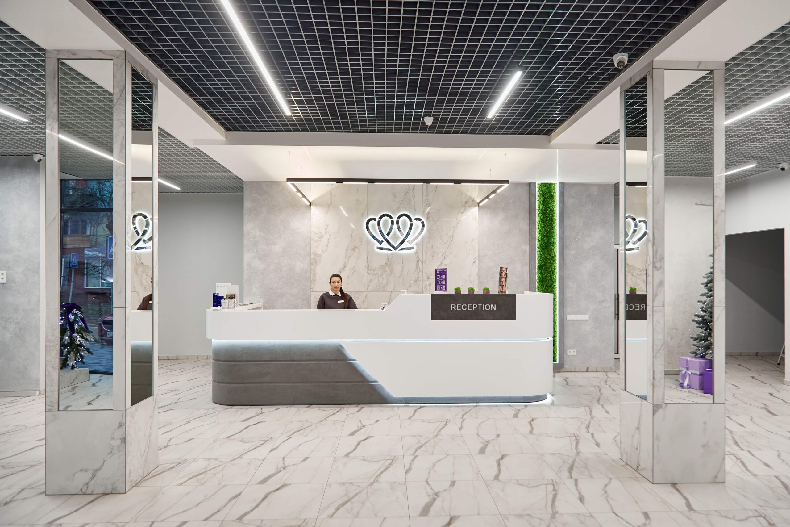
📍 Lviv, Ukraine
A dental clinic in Lviv with a separate doctor for each field: oral surgery and implants, prosthodontics, restorative dentistry, endodontics under a microscope, periodontics, orthodontics and paediatric dentistry.
No. A deeply impacted tooth with no symptoms, no cyst and no effect on its neighbours in an adult patient is left under observation with an examination and an X-ray once every year or two. Removing all unerupted teeth preventively has not been shown to be beneficial. It is a different matter when the tooth presses on the root of a neighbour, inflammation flares up repeatedly under the gum, a follicular cyst is visible on the X-ray or the tooth gets in the way of an orthodontic plan: then delay only makes the eventual operation bigger.
At MediUkraine's partner clinic, surgical removal of a third molar starts from USD 52, a CT scan of both jaws costs from USD 15, computer-controlled anaesthesia from USD 13, and drug sedation from USD 33 per 30 minutes. The starting price applies to a more straightforward case; deep impaction with bone removal and sectioning of the tooth into fragments costs more, and the surgeon names the figure after a three-dimensional scan. Prices are a guide; the clinic sets the final figure.
To see what a flat X-ray does not show: the real position of the unerupted tooth, the number and shape of its roots, the thickness of bone over the crown and the distance to neighbouring structures. In the lower jaw that means the mandibular canal with the nerve responsible for sensation in the lip and chin, in the upper jaw the floor of the maxillary sinus, and for a canine also the condition of the lateral incisor's roots. The access technique, the length of the operation and the likelihood of paraesthesia depend on these millimetres.
Usually it is brought into the arch. The canine carries chewing load and shapes the smile line, so with a moderate tilt and a sound tooth, surgical exposure of the crown with attachment of a chain is planned, followed by orthodontic traction lasting months. Removing the tooth is discussed when it is ankylosed, lies almost horizontally, has a sharply curved root or has already caused resorption of the neighbours' roots. The choice is made by the surgeon and the orthodontist together, based on the CT scan and the patient's age.
It is a loss or change of sensation in the lower lip, chin or half of the tongue after surgery in the lower jaw. The cause is injury to, or swelling around, the nerve that runs close to the roots of the tooth. It feels like persistent numbness, as if the anaesthetic is not wearing off. In most cases sensation returns within a few weeks or months; persistent loss is rare. Numbness that lasts more than a day after the operation should be reported to the surgeon, who will assess the situation and prescribe treatment if needed.
The operation itself is done under anaesthesia, and the patient feels no pain: you will feel pressure and movement, but not pain. For lower teeth a nerve block is given, for upper teeth infiltration, and the surgeon begins only after checking that the area is numb. If the anaesthetic starts to wear off during a long procedure, more is given. If you find dental treatment stressful, drug sedation is available; you will then need someone to take you home and must not drive that day.
Swelling builds up until day 2–3 and then subsides; at the same time the limited mouth opening gradually passes. Most people return to their usual routine within 5–10 days, and sutures are removed on day 7–10. The soft tissue closes in roughly 2–3 weeks, and the bone remodels for another 3–6 months, a stage the patient no longer feels. After deep impaction recovery takes longer than after a simple extraction, because the scope of the procedure is greater. Ask your doctor for the exact timescales in your case.
Tell us what you need: we will suggest a clinic and a doctor and send you a cost estimate.
Request a consultation →