| Service | 🇺🇦 Ukraine | 🇨🇿 Czech Republic | 🇹🇷 Turkey |
|---|---|---|---|
| Dentist consultation | from USD 8 | from USD 38 | from USD 34 |
| Online consultation | from USD 8 | — | — |
| Consultation with CT scan | from USD 20 | from USD 114 | from USD 92 |
| Treatment plan | from USD 14 | — | — |
Prices in the “Ukraine” column come from MediUkraine’s partner clinics. The Czech Republic and Turkey columns show the lower bounds of prices published by clinics there (for Turkey, also the Turkish Dental Association’s recommended 2026 tariff), collected in August 2026 and converted to US dollars at the rate on the day of collection. A dash means we could not find a comparable published price. In any country, the final cost is set by the clinic after an examination.
What sets a first appointment apart from a visit to "look at one tooth" is its scope. The dentist examines all the teeth, the lining of the cheeks and lips, the tongue, the floor of the mouth and the palate, and assesses the gums around each tooth, the condition of old fillings and crowns, and the way the teeth meet. People usually come in because one tooth hurts, and the examination finds two or three other problems that do not hurt yet and so have gone unnoticed.
The aim is not to list cavities but to understand the state of the mouth as a whole and plan the order of treatment. The same crown can be the end goal or a first step: if there is inflammation under it and the neighbouring tooth is loose because of periodontitis, the order of procedures changes, and with it the quote. That is why the examination ends with a plan, not a list of diagnoses.
A routine check-up is worthwhile every 6–12 months, even when nothing is bothering you: caries on the contact surfaces and early periodontitis produce no symptoms for years. People book an unscheduled visit for pain, bleeding gums, a chipped tooth, a lost filling, bad breath or loose teeth, and also before prosthetic work, implants and orthodontic treatment, since these start only once all existing dental problems have been treated.
Sometimes a routine examination is not what you need right now. Throbbing pain that keeps you awake, swelling of the cheek or gum, a raised temperature, or an injury to a tooth with displacement or a broken fragment need an emergency appointment the same day, not a booking on a convenient date. An examination is not treatment either: it does not relieve pain and is no substitute for either treatment or professional cleaning. And a remote consultation, useful as it is, cannot provide a final diagnosis.
MediUkraine is a treatment search and coordination service, not a clinic. We choose the dental clinic and a dentist with the right specialism, agree the date of the visit and support you throughout, including if you are travelling from abroad. The diagnosis and the scope of treatment are decided by the dentist after an in-person examination.

Travel. Ukrainian airspace is closed, so there are no commercial flights. Patients reach Lviv overland, most often from Poland: Rzeszów and Kraków airports are the usual entry points, with a train, bus or private transfer onward to the border and on to the city. Lviv is roughly 70 km from the Polish border, the shortest crossing of any Ukrainian city.
Visas. Citizens of the EU, the United Kingdom and the United States do not need a visa for short stays in Ukraine. Rules depend on your citizenship, so check your own before booking.
Safety, plainly. Ukraine is at war. Air raid alerts occur in Lviv, and an appointment can be interrupted by one. Several governments advise against travel to Ukraine, and while that advice stands, ordinary travel and medical insurance is often void — confirm your cover with your insurer before you commit to anything.
Language. MediUkraine handles correspondence, scheduling, the written treatment plan and the quote in English. The clinic's team communicates with international patients in English.
Payment. You pay the clinic directly. Our coordination service is free of charge for you.
More on travel, safety and insurance: Safety and travel.
Talking through your symptoms is not a formality but the first diagnostic step. The nature of the pain narrows down the possible causes even before the examination: a brief pain from cold that passes within seconds behaves differently from throbbing night pain, and pain when biting on a specific tooth differs from diffuse pain you cannot pinpoint. The dentist asks when it started, what preceded it, whether you took painkillers and whether they helped, how this tooth was treated before and whether there were complications after previous procedures.
The examination begins outside the mouth: the dentist checks the symmetry of the face, feels the lymph nodes under the jaw and in the neck, and assesses how wide the mouth opens and whether there are sounds in the temporomandibular joint. Then the lining of the cheeks and lips, all sides of the tongue, the floor of the mouth, the palate and the tonsils are examined. This takes a minute or two, but it doubles as oral cancer screening: most growths in the mouth are painless at an early stage and are found mainly at routine examinations.
Probing is a systematic check of each tooth with a mirror and a probe on a dried surface. The dentist looks for cavities, chalky spots of early demineralisation, enamel cracks, chips, overhanging margins and gaps under old fillings, and the condition of crown margins. The method has a limit: the probe should not be pressed into a fissure, because this is an unreliable way to detect caries and at the same time risks damaging enamel in which demineralisation is still reversible. That is why probing is combined with drying, good light, magnification and, where available, transillumination of the tooth, while the contact surfaces between the teeth are assessed on X-rays.
Pulp vitality tests are needed when a tooth is bothering you or has a deep old filling. A cold test shows whether the nerve responds and how quickly the response fades; percussion (tapping on the tooth) points to inflammation at the root tip; palpation of the gum over the root reveals tenderness and swelling. These simple tests, together with an X-ray, distinguish reversible pulp inflammation, which is treated with a filling, from irreversible inflammation, which needs root canal treatment. A mistake at this stage can cost a tooth, so the dentist compares the reaction of the problem tooth with that of its neighbours and of the matching tooth on the opposite side.
The periodontal chart is what distinguishes a thorough examination from a cursory one. Using a graduated probe, the dentist measures the depth of the gum crevice at six points around each tooth and records the results. A crevice 1–3 mm deep without bleeding is considered healthy, 4–5 mm means a pocket, and 6 mm or more indicates deep disease. Bleeding on probing, gum recession, tooth mobility, furcation involvement (where the roots divide) and the amount of plaque are recorded at the same time.
These numbers add up to a diagnosis. Based on pocket depth, loss of attachment of the gum to the tooth and the picture on the X-ray, periodontitis is described by a stage, reflecting its severity and the amount of bone lost, and a grade, reflecting the rate of progression; its extent is assessed separately, as localised or generalised. Bleeding at more than a tenth of the measured points means active inflammation, even if the pockets are shallow. But what matters most for you is this: the chart is a set of recorded numbers that can be compared six months later to see whether things are better or worse, instead of relying on the phrase "the gums are a little inflamed".
X-rays cover what cannot be seen in the mirror. A periapical X-ray shows one or two teeth together with the root and the surrounding bone: it is used to assess the root canals, a focus of inflammation at the root tip and the quality of earlier root fillings. Bitewing X-rays of the back teeth show caries on the contact surfaces and the bone level between the teeth, areas that cannot be examined any other way. A panoramic X-ray (orthopantomogram) gives an overview of both jaws with the joints and wisdom teeth, but distorts scale and is unsuitable for precise measurements.
Cone beam CT is not needed by everyone or every time. It is ordered for specific indications: implant planning, complex root canal anatomy, suspected root fracture, an impacted wisdom tooth close to the nerve canal, cysts and long-standing inflammation. The three-dimensional image shows the volume and density of bone and the position of the mandibular canal and the maxillary sinuses. A CT scan should not be taken just in case: its radiation dose is higher than that of an ordinary X-ray, so the method is chosen to fit the clinical question. An intraoral camera and a photo record involve no radiation at all and also give a baseline for comparison at later examinations.
The treatment plan is the outcome of the appointment and the most important document you take away. It turns the findings into a sequence: an emergency stage if there is pain or inflammation; hygiene and treatment of existing problems; periodontal treatment with re-assessment; root canal treatment; and only then prosthetics, implants or orthodontics. Each stage comes with an approximate cost, number of visits and timeline, and where there are options (an inlay instead of a large filling, a crown on your own tooth instead of extraction and an implant), the dentist describes the alternatives and their limitations. At this appointment the bite is assessed visually; if the plan involves extensive prosthetic work, a separate functional examination is ordered.
An online consultation uses the same information, gathered in advance. For it to be useful, send the dentist a panoramic X-ray or CT scan as the original DICOM file rather than a photograph of a screen; periapical X-rays, if you have them; photographs of the teeth from several angles, the smile, the closed teeth from the front and the sides, the upper and lower arches from above; discharge summaries and descriptions of work done; and a list of medicines and chronic illnesses. From these, the dentist assesses the likely scope of work, the order of the stages and the approximate cost, warns of possible difficulties and estimates how many days treatment will take. What a remote consultation cannot do is make a final diagnosis: pocket depth, tooth mobility, the condition of old filling margins and the pulp's response can be checked only with an instrument in the mouth. That is why the preliminary plan is refined after an in-person examination, and sometimes during treatment, when the dentist removes an old filling and sees the true extent of the damage. You are told about this possibility honestly at the start, not after the fact.
Straight after the examination the dentist goes through the findings with you on screen, showing the X-rays, the findings in the photographs and the figures of the periodontal chart. You receive the summary and the X-ray files the same day. If the work is extensive and involves several specialists, the written plan with a quote is ready within 1–3 working days, because the prosthodontist, surgeon and periodontist agree it between them.
How soon the next steps happen depends on how urgent the situation is. Pain and inflammation are dealt with the same day or the next. Uncomplicated cavities take one visit each, and treating all existing problems usually takes 2–4 visits over two to four weeks. Root canal treatment takes one or two visits per tooth, sometimes a few days apart.
The periodontal stage is assessed separately and later than the others. After professional cleaning and removal of deposits below the gum line, the periodontal chart is repeated after 6–8 weeks; before then the tissues have not had time to respond. Comparing the two charts, not how you feel, shows whether the pockets have shrunk and the bleeding has stopped, and whether the next stage is needed.
Routine check-ups are scheduled every 6–12 months depending on risk: more often with active caries, a dry mouth or periodontitis, less often when the situation is stable. Bitewing X-rays are repeated at an interval the dentist sets individually, and a panoramic image of both jaws is taken not on a schedule but when there is an indication for it.
The plan can change once work is under way, and this is a normal part of the process. When removing an old filling, the dentist sometimes finds more extensive damage or a crack that did not show on the X-ray; occasionally a tooth that was meant to be saved turns out to be beyond saving. In that case work stops, the situation is explained and any change to the quote is agreed before continuing, not afterwards.
If you live abroad, your documents are prepared so that treatment can continue at home: a summary of the findings, the periodontal chart and the X-ray files. An interim check after you return can be done remotely from photographs and your description of how you feel, but measuring pockets and checking how well fillings fit has to be done in person, at home or on your next trip.
Step 1. Your symptoms and history. The dentist finds out what is bothering you and for how long, what triggers the pain and whether it wakes you at night, how your teeth were treated before, and whether there were complications after anaesthesia or extractions. The dentist also asks about chronic illnesses, medicines, allergies and pregnancy. This part may seem optional, but it determines which tests and X-rays will be needed next.
Step 2. Examination and probing. First the face, lymph nodes and mouth opening are checked, then the lining of the cheeks, the tongue and the palate, and only then the teeth. Each tooth is examined with a mirror and a probe on a dried surface for fillings, crowns, cracks and early enamel changes. If needed, a cold test and percussion show the condition of the nerve.
In total, the first appointment takes about 30–40 minutes. After talking with you and examining your mouth, the dentist checks the teeth with a probe, makes a periodontal chart measuring the pockets at six points around each tooth, takes X-rays where indicated, shows the findings on the screen and ends the appointment with a treatment plan with stages and a quote. No anaesthesia, hospital stay or recovery period is needed: straight after the visit you can eat, talk and return to your usual activities.
Stage zero is emergency care, and not everyone needs it. If the examination finds acute pain, an abscess or swelling, the inflammation is dealt with first and the rest is planned afterwards. This stage takes one or two visits and comes before everything else, because neither prosthetic work nor orthodontics can start while acute inflammation is present.
The first stage is hygiene and treatment of existing problems. Deposits are removed, you are shown how to clean your teeth properly, caries is treated and defective fillings are replaced. This usually takes 2–4 visits over two to four weeks and is necessary even if you came for crowns: in a mouth with inflamed gums and active caries no prosthetic work lasts long.
The second stage is periodontal and endodontic. Pockets are cleaned under local anaesthesia, root canals are treated or retreated, and teeth beyond saving are extracted. After 6–8 weeks the periodontal chart is repeated and compared with the first: this decides whether you can move on to the next stage or need gum surgery.
The third stage is restoration: crowns, inlays, veneers, implant-supported prosthetics, orthodontic treatment. This is the longest stage. Bone healing after implant placement takes roughly 3–6 months, orthodontic treatment from a few months to two years, and temporary restorations are worn for several weeks before the permanent ones.
The fourth stage is maintenance, and without it the results of the previous three gradually wear off. It means a check-up and professional cleaning every 6–12 months, monitoring of periodontal measurements and, if needed, a night guard. For patients from abroad the cycle is split into two or three trips: the first for treating existing problems and preparation, the second for the main work, the third for completion, with interim checks done remotely.
The examination itself is non-invasive: no tissue is damaged, no anaesthesia is used and there is no recovery period. Pocket measurement may cause moderate discomfort, especially if the gums are inflamed, and some bleeding on probing. Bleeding here is not a complication but a diagnostic sign: a healthy gum does not bleed when touched with a probe. If the gums are very tender, the dentist uses a topical anaesthetic or postpones the measurement until the acute inflammation has settled.
X-rays involve radiation, and it helps to see this in figures. The effective dose of a digital periapical X-ray is measured in single microsieverts, of a panoramic image of both jaws in tens, and of cone beam CT from several tens to several hundred microsieverts depending on the machine and the field of view. For comparison, natural background radiation gives each of us a few microsieverts a day. X-rays are taken where indicated, with a protective apron and, for intraoral X-rays, a thyroid collar, and for pregnant women they are taken only when they cannot be avoided, after discussion with the doctor managing the pregnancy.
Infection control matters too. Examination instruments are sterilised in an autoclave, handpieces are reprocessed after each patient, and disposable gloves, masks, bibs, saliva ejectors and protective barriers on the dental unit buttons are used. The X-ray sensor is covered with a disposable sleeve. You are entitled to ask how the clinic handles sterilisation and to get a specific answer.
General health conditions and medicines need particular attention. Anticoagulants and antiplatelet drugs increase bleeding during probing and extractions, but you must not stop them yourself before the visit: that decision belongs to the doctor who prescribed them. Bisphosphonates (especially intravenous) and denosumab injections are associated with a risk of osteonecrosis of the jaw after extractions and implant placement, so tell the dentist about them before planning begins. Patients with certain heart defects, prosthetic heart valves or a history of infective endocarditis are, where indicated, given antibiotic prophylaxis before procedures that involve bleeding. Uncontrolled diabetes slows healing and worsens the course of periodontitis.
Remote consultations have clear limits, and it is worth knowing them before you pay. Photographs and X-rays cannot show pocket depth, tooth mobility, the nerve's response or how tightly a filling fits. So an online consultation gives a guide to the scope and order of work, not a diagnosis and not a final quote. If an exact plan is promised remotely without an examination, that is a reason to be wary: the plan will most likely change, and not in the patient's favour.
The clinics MediUkraine works with hold a licence for medical practice from the Ministry of Health of Ukraine. You receive a written summary and the X-ray files, so you can always get a second opinion: the documents can be shown to another specialist, and if needed we arrange a repeat appointment at another partner clinic. Only a dentist who has examined you can predict the outcome of your treatment, and you decide on the scope of treatment yourself, without time pressure.
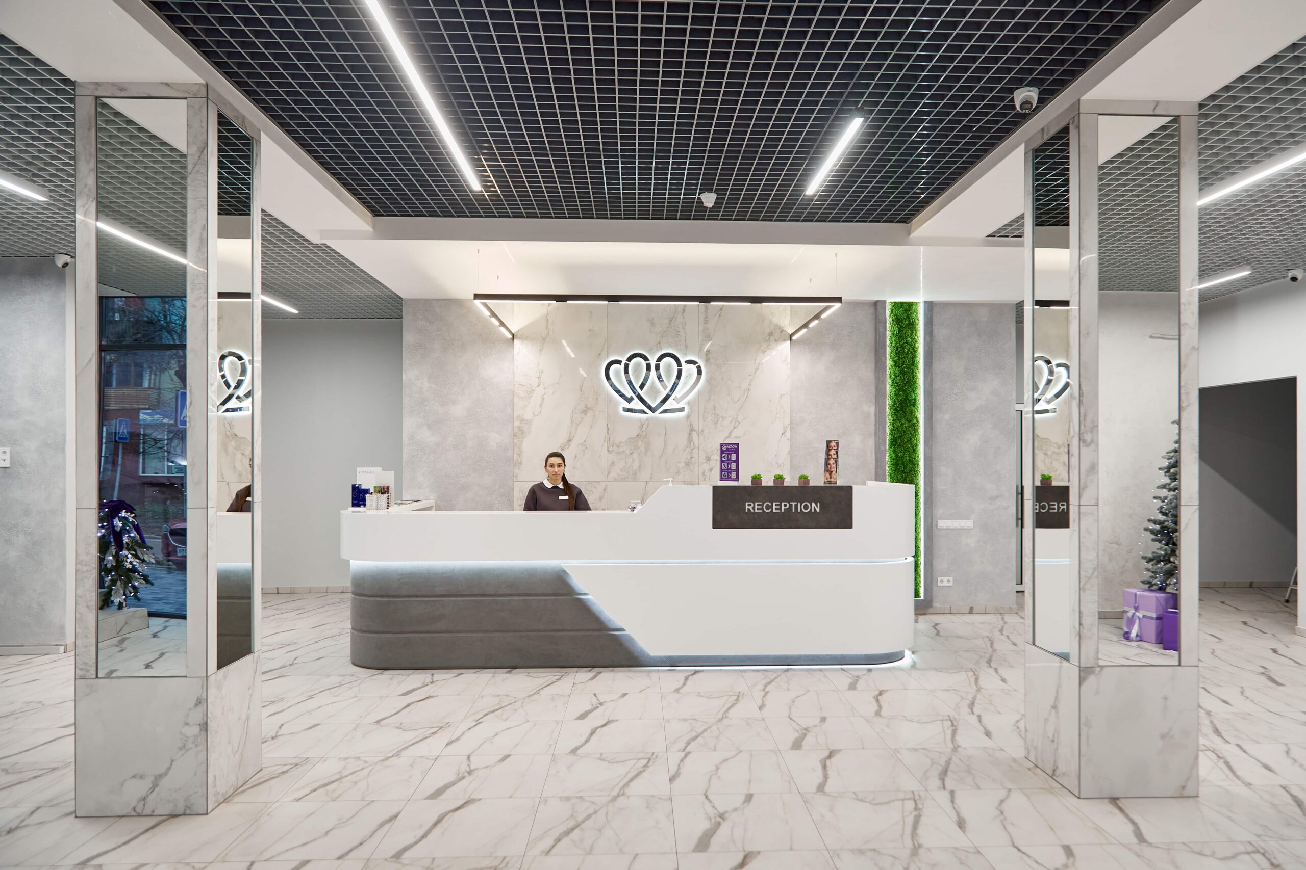
📍 Lviv, Ukraine
A dental clinic in Lviv with a separate doctor for each field: oral surgery and implants, prosthodontics, restorative dentistry, endodontics under a microscope, periodontics, orthodontics and paediatric dentistry.
A discussion of your symptoms and history, an examination of the face, lymph nodes and oral lining, a systematic examination of all the teeth with a mirror and a probe on a dried surface and, if needed, tests of the nerve's response. Then the dentist makes a periodontal chart: the depth of the gum crevice is measured at six points around each tooth, and bleeding, recession and mobility are recorded. X-rays are taken where indicated. The appointment ends with a review of the findings on screen and a treatment plan with stages, timelines and a quote.
At the clinics MediUkraine works with, a first appointment costs from USD 8 and usually includes the examination, probing and a discussion of the findings. A remote appointment costs the same, from USD 8. A consultation with a CT scan is charged separately, from USD 20 and a written treatment plan with stages and a quote from USD 14. Periapical X-rays, a panoramic image of the jaws, professional cleaning and the treatment itself are separate items. The clinic confirms the final cost after an in-person examination.
It is a chart in which the dentist records the depth of the gum crevice at six points around each tooth, as well as bleeding on probing, gum recession, mobility and plaque. A crevice of 1–3 mm without bleeding is considered healthy, 4–5 mm means a pocket, and 6 mm or more indicates deep disease. The chart does two things: it turns a vague phrase about inflamed gums into specific numbers, and it provides a baseline. The measurements are repeated 6–8 weeks after treatment and compared, so you can see whether treatment has worked.
Not always, but without them the picture is often incomplete: caries on the contact surfaces between the teeth, the condition of the root canals and the bone level cannot be seen in a mirror. The dentist decides which images are needed: for some patients one periapical X-ray is enough, others need bitewing X-rays of the back teeth or a panoramic image of both jaws. CT is ordered selectively, for implant planning, complex canal anatomy or impacted wisdom teeth. If you have recent X-rays, bring them with you: there is no point in repeating X-rays unnecessarily.
Previous X-rays on a disc or as files, discharge summaries and contracts describing work done, certificates for implants that have been placed, removable dentures, splints and retainers. Be sure to bring a list of the medicines you take regularly, with names and doses, as well as information about allergies, chronic illnesses, pregnancy and past surgery. It helps to write down beforehand what is bothering you and for how long, because details are easily forgotten in the chair. Book professional cleaning after the examination, not the day before.
It is a video call based on materials you send in advance: a panoramic X-ray of the jaws or a CT scan as the original DICOM file, periapical X-rays, photographs of the teeth from several angles, the smile, the closed teeth from the front and the sides, the upper and lower arches from above, as well as discharge summaries and a list of medicines. The dentist assesses the likely scope of work, the order of the stages, the approximate cost and how many days the trip will take. It cannot provide a final diagnosis: pocket depth and the nerve's response can be checked only with an instrument in the mouth.
Often, yes, and for patients from other cities clinics deliberately plan the visit with time to spare. On the day of the examination it is usually realistic to relieve acute pain, treat one or two uncomplicated cavities, start root canal treatment or have professional cleaning. Major work (prosthetics, implants, orthodontic treatment) does not start the same day: treating existing problems, reviewing the X-rays and agreeing a plan come first. If you are short of time, mention it when booking, and the schedule will be planned so that the first stage fits into your trip.
Tell us what you need: we will suggest a clinic and a doctor and send you a cost estimate.
Request a consultation →