| Service | 🇺🇦 Ukraine | 🇨🇿 Czech Republic | 🇹🇷 Turkey |
|---|---|---|---|
| Gum (soft tissue) grafting | from USD 73 | from USD 239 | from USD 241 |
| Curettage (1 tooth) | from USD 13 | from USD 14 | from USD 39 |
| Periodontist consultation | from USD 18 | — | — |
Prices in the “Ukraine” column come from MediUkraine’s partner clinics. The Czech Republic and Turkey columns show the lower bounds of prices published by clinics there (for Turkey, also the Turkish Dental Association’s recommended 2026 tariff), collected in August 2026 and converted to US dollars at the rate on the day of collection. A dash means we could not find a comparable published price. In any country, the final cost is set by the clinic after an examination.
Recession is when the gum margin recedes and exposes part of the tooth root. The reference point is the cemento-enamel junction: normally the gum covers it, but with recession 1–5 mm or more of root shows above the gum. The root is covered not by enamel but by cementum, so it looks darker, wears faster and is more sensitive to cold.
There are usually several causes at once: a thin gum phenotype and a narrow band of keratinised gum, missing bone over the outer surface of the root, hard brushing with horizontal strokes and a hard brush, a high frenulum or muscle attachments, a tooth moved outside the bone during orthodontic treatment, chronic gum inflammation, or a lip or tongue piercing. The cause determines not only the technique but also whether the result will last.
The surgery is not only about appearance. An exposed root is sensitive to cold, sweet things and touch, is prone to decay at the gum line and is harder to clean, and an area without firm gum is easily injured by the toothbrush and becomes inflamed. Another reason is preparation for orthodontic treatment or crowns: with thin tissue, moving a tooth or placing a crown margin often makes a recession worse, so the gum is thickened in advance rather than after a problem appears.
The doctor decides on treatment after taking measurements, and not every recession needs surgery. A shallow defect that is not sensitive or getting worse is monitored: it is photographed and measured every six months and, above all, brushing technique is corrected. Surgery is not done with active inflammation and poor hygiene, uncontrolled periodontitis, or when the bone and papilla between the teeth have already been lost: in that case the root cannot be covered, and this needs to be discussed before surgery, not after.
MediUkraine is a treatment search and coordination service, not a clinic. We choose the clinic and a doctor with the right specialism, arrange the visit dates and support you throughout, including if you are travelling from abroad. The diagnosis, indications and treatment plan are decided by the doctor after an in-person examination.

Travel. Ukrainian airspace is closed, so there are no commercial flights. Patients reach Lviv overland, most often from Poland: Rzeszów and Kraków airports are the usual entry points, with a train, bus or private transfer onward to the border and on to the city. Lviv is roughly 70 km from the Polish border, the shortest crossing of any Ukrainian city.
Visas. Citizens of the EU, the United Kingdom and the United States do not need a visa for short stays in Ukraine. Rules depend on your citizenship, so check your own before booking.
Safety, plainly. Ukraine is at war. Air raid alerts occur in Lviv, and an appointment can be interrupted by one. Several governments advise against travel to Ukraine, and while that advice stands, ordinary travel and medical insurance is often void — confirm your cover with your insurer before you commit to anything.
Language. MediUkraine handles correspondence, scheduling, the written treatment plan and the quote in English. The clinic's team communicates with international patients in English.
Payment. You pay the clinic directly. Our coordination service is free of charge for you.
More on travel, safety and insurance: Safety and travel.
How much of the root can be covered depends not on the technique but on the tissue between the teeth, so everything starts with classification. The Miller classification has four classes: I, the recession does not reach the mucogingival junction, and the bone and papilla between the teeth are preserved; II, the recession reaches or extends beyond the junction, but there is no loss between the teeth; III, there is already loss of bone or soft tissue between the teeth, or the tooth is out of position; IV, marked tissue loss all around the tooth. In Classes I and II full root coverage is predictable, in Class III partial coverage is usually the realistic goal, and in Class IV the root cannot be covered.
The later Cairo classification (RT1, RT2, RT3) does the same thing more simply, based on the attachment level between the teeth, which the doctor measures with a probe rather than judging by eye. RT1 means no attachment loss between the teeth, RT2 means loss between the teeth no greater than on the outer surface, and RT3 means greater loss between the teeth. The practical point is the same: the height of the papilla between the teeth is the ceiling, and the gum margin will not rise above it after surgery.
The coronally advanced flap is the basic operation for covering recession. The gum over the defect is lifted, released and moved up above the cemento-enamel junction, where it is secured with microsutures. The method works where a band of firm keratinised gum remains below the recession; on its own it adds almost no thickness, so it is used without a graft only with a thick phenotype and shallow defects.
The tunnel technique needs no vertical incisions: special instruments are passed through the gum crevice to create a tunnel under the papillae, and the flap stays in one piece. The blood supply suffers less and there are no incision scars, which is why it is often chosen in the smile zone and for several neighbouring recessions. The tunnel is almost always combined with a graft, which is slid into the space and drawn up with sutures together with the gum margin.
A connective tissue graft from the palate remains the gold standard against which other materials are judged. A piece about 1–1.5 mm thick is taken from between the canine and the first molar, a few millimetres from the gum margin; the palatine artery runs deeper, so the soft-tissue thickness is assessed in advance. The donor wound is closed with sutures, a collagen sponge and a protective plate, and the surface heals over in about 2–3 weeks. The graft thickens the gum and widens the band of keratinised tissue, and this is what determines whether the gum margin will hold a year later.
A free gingival graft solves a different problem: not covering the root but creating a band of firm gum where there is none. It is taken from the palate with its surface layer and stitched onto a prepared site. The method is predictable, but the grafted area usually differs in colour and texture from the surrounding gum, so it is used sparingly in visible areas and more often in the lower jaw, around implants and before crowns or bridges.
Substitutes for your own tissue avoid a second wound. A xenogeneic collagen matrix or an acellular dermal matrix causes less discomfort and is not limited by the size of the palate; studies show they come close to a graft for root coverage but fall short of it for gain in tissue thickness. Enamel matrix proteins applied to the cleaned root surface are also used as an adjunct. The choice of material depends on the area of the recessions, the thickness of the palate and how much of a gain in gum volume is needed.
Sometimes working on the gum alone is not enough. A laterally positioned flap is used for a single defect when the neighbouring area has enough tissue to donate; vestibuloplasty deepens the vestibule if soft-tissue tension keeps pulling the gum margin down; a high or thick frenulum needs separate surgery, which has its own page in our catalogue. With a wedge-shaped defect or wear at the neck of the tooth, surgery is combined with a filling: the part of the defect nearer the crown is restored with composite, and the part nearer the root tip is covered with your own tissue.
Gummy smile correction is a related branch of the same surgery. Excessive gum display is generally defined as roughly 3–4 mm or more in a broad smile, and the causes vary: incomplete eruption, where the gum covers part of the crown, excessive vertical growth of the upper jaw, or a short or overactive upper lip. For incomplete eruption, gingivoplasty with clinical crown lengthening is performed; the attachment zone above the bone crest, about 3 mm, must always be respected, otherwise the gum margin will grow back or chronic inflammation will develop.
When the cause is not in the gums, gum surgery will not help. For an overactive lip, lip repositioning surgery or temporary muscle relaxation with botulinum toxin may be considered; the effect lasts about 4–6 months and needs repeating. The skeletal form, excessive vertical growth of the upper jaw, is treated with orthognathic surgery together with a maxillofacial surgeon, while over-erupted front teeth are corrected orthodontically. So the first step with a gummy smile is finding the cause, not booking a gingivoplasty.
For the first two or three days expect swelling, possibly bruising on the cheek and an ache that ordinary painkillers relieve. On the first day apply cold to the cheek, and sleep with your head raised on a high pillow. The donor site on the palate is usually more uncomfortable than the grafted area itself, especially when eating.
Do not brush the treated area for 2–4 weeks: pressure can shift the flap and graft, which in the first days are held only by stitches and a thin layer of fibrin. Instead, use 0.12% chlorhexidine rinses or applications twice a day. Brush the rest of your teeth as usual, and go back to a normal toothbrush on the treated area only when the doctor says so, and without pressure.
For a week, stick to soft, cool food (nothing hot, spicy or hard) and avoid sport, saunas and straining or bending over. Do not pull your lip back to look at the surgical site: this is the most common way the flap gets displaced. Stitches come out on day 10–14, sometimes in two stages: first from the palate, then from the grafted area.
There is no point judging how the gum looks as soon as the stitches come out. Swelling and redness subside over 1–2 months, colour and texture even out more slowly, and the final contour forms after 3–6 months. That is when follow-up photos are taken from the same angles and compared with the pre-surgery ones.
The result is measured in millimetres, not judged by eye: the doctor re-measures the recession depth and the width of keratinised gum. In Miller Classes I and II full root coverage is predictable; in Class III partial coverage is agreed as the goal before surgery. In some patients the gum margin creeps a little further over the root over the following months, adding 0.5–1 mm.
Sensitivity usually decreases once the root is covered, although in the first weeks it can even get worse because of the procedure itself. After that, the result depends on two things: careful daily hygiene and removing the cause of the recession. Check-ups with professional cleaning are scheduled every 3–6 months, and only a doctor can predict the result in your case, after an examination.
Step 1. Consultation and measurements. The doctor examines every area with exposed roots, uses a probe to measure the depth of each defect, the width of keratinised gum and the tissue level between the teeth, and assesses gum thickness and brushing technique. These measurements determine the class of recession and so how much root coverage can be expected. Photos and, if needed, X-rays are taken at the same visit.
Step 2. Preparing the tissue. Professional cleaning, curettage where there are pockets, replacement of overhanging fillings at the gum line and training in gentle brushing. Surgery is scheduled once the gums no longer bleed on probing: a graft takes less well on inflamed tissue, so this step is not cut short to get an earlier date.
In total, it takes 3–6 months from the first consultation to the final assessment of the contour, although the active treatment takes a single day of surgery. Between the measurements and preparatory cleaning at the start and the follow-up check-ups at the end come the surgery itself (a coronally advanced flap or tunnel technique, with a palatal graft if needed) and two weeks until the stitches come out. How many areas need separate procedures becomes clear at the first examination, once the measurements are taken.
Treatment has four parts: preparation, surgery, healing and maintenance. Surgery is the shortest, one visit in a process lasting weeks or months. The result depends less on the day of surgery than on what happens before and after it.
Preparation usually takes 2–4 weeks. During this time inflammation is brought under control, cleaning and, if needed, curettage are done, brushing technique is corrected and overhanging fillings are replaced. If you smoke, this is also the time to stop, ideally keeping it up after the procedure as well.
Surgery is done one area at a time, not on the whole mouth at once. One visit usually covers recessions on one to four neighbouring teeth: more is harder to tolerate, and the donor area on the palate is limited. The next area is planned no sooner than 6–8 weeks later, once the first has healed.
Healing has its own timetable too. Stitches come out on day 10–14, the palate heals over within 2–3 weeks, swelling subsides over 1–2 months, and the contour and colour of the tissue settle after 3–6 months. That is when follow-up photos and repeat measurements are taken, not before.
After that comes maintenance. Check-ups with professional cleaning are scheduled every 3–6 months to review brushing technique and the gum margin. If orthodontic treatment or crowns are planned, the order is agreed in advance: thin gum is usually thickened before the teeth start to move, while a crown margin is shaped only after the gum contour has settled.
Expected after-effects include swelling, bruising on the cheek, aching for the first two or three days and a sore palate that makes eating difficult for one to two weeks. These are not complications but part of normal healing, and you will be told about them before surgery, not after.
The main specific risk is partial take of the graft or displacement of the flap. In the first days the grafted tissue survives on diffusion, so tension in the flap, pressure from a toothbrush or the lip, smoking and heavy physical exertion can cost part of the result, leaving part of the root exposed. A repeat procedure is then planned no sooner than several months later, once the tissue has fully recovered.
The donor site has its own risks. The most common is bleeding in the first hours; less common are exposed bone under the stitches and slower healing; rarer still is a temporary change in sensation in the palate. Soft-tissue thickness and the course of the palatine artery are assessed beforehand so the surgeon does not go deeper than the anatomy allows; a protective plate and a collagen sponge noticeably reduce discomfort in the first days.
The recession can come back if its cause remains. Returning to hard, horizontal brushing, smoking, a lip piercing, pull from the frenulum or muscle attachments, moving a tooth outside the bone during orthodontic treatment, untreated inflammation: all of these gradually undo the result. That is why correcting brushing technique and monitoring hygiene are part of the treatment itself, not optional advice.
Planned surgery is postponed with active gum inflammation and poor hygiene, acute infections, uncontrolled diabetes and severe clotting disorders; during pregnancy, planned procedures are usually deferred. Particular caution is needed for patients taking bisphosphonates or denosumab or who have had radiotherapy to the head and neck: their risk of jaw osteonecrosis is higher, so whether surgery is advisable is agreed with the doctor treating the underlying condition. Anticoagulants do not rule out the procedure but require planning, and you must not stop them on your own.
A word about expectations. Surgery covers the root but does not restore a papilla lost between the teeth: "black triangles" between the necks of the teeth usually remain after surgery, and this is discussed in advance. The clinics MediUkraine works with hold a Ukrainian Ministry of Health licence for medical practice, use sterile instruments and obtain written informed consent listing the possible complications. The surgical report and photos stay with you, so you can get a second opinion from another specialist if needed.
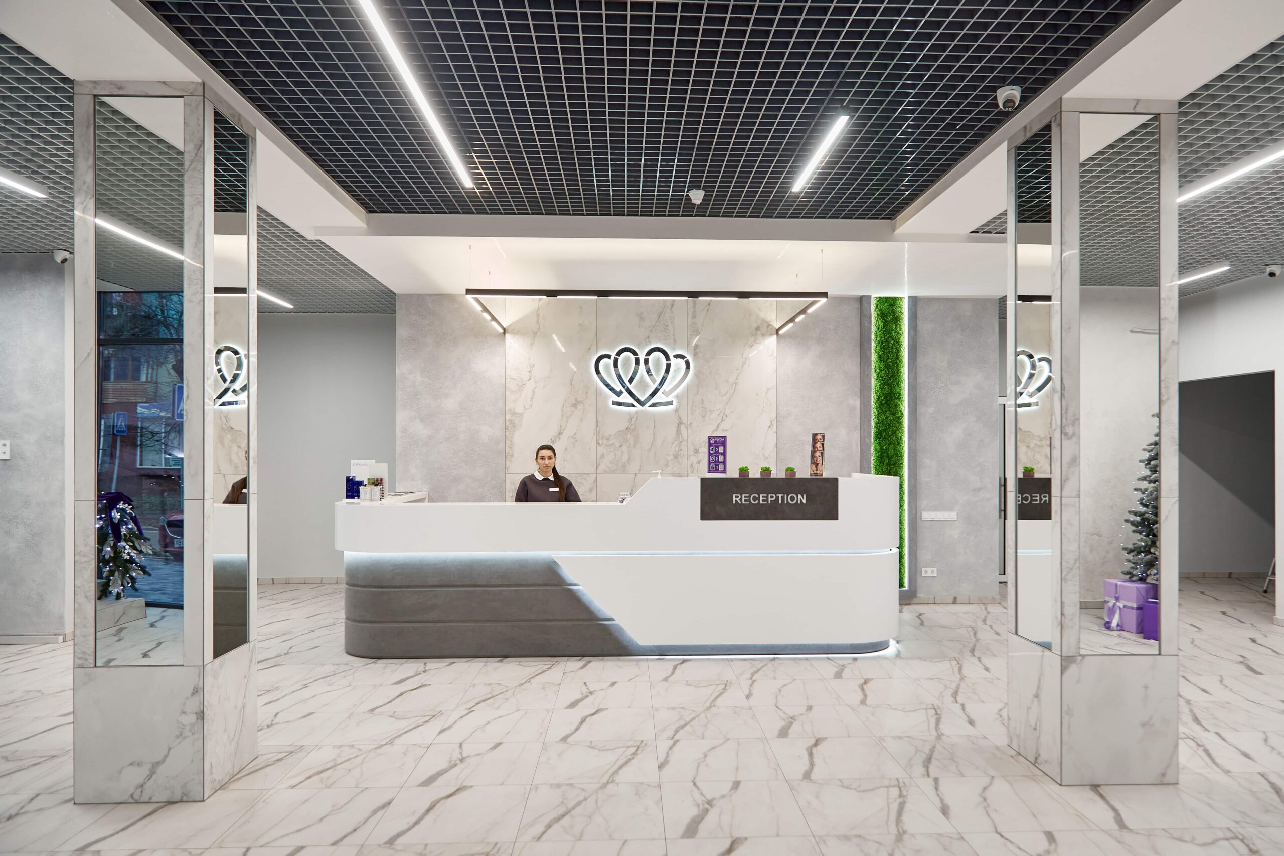
📍 Lviv, Ukraine
A dental clinic in Lviv with a separate doctor for each field: oral surgery and implants, prosthodontics, restorative dentistry, endodontics under a microscope, periodontics, orthodontics and paediatric dentistry.
It depends on the tissue between the teeth. Under the Miller classification, in Classes I and II, where the bone and papilla between the teeth are intact, full root coverage is predictable. In Class III, where some has already been lost, partial coverage is expected, and in Class IV the root cannot be covered: surgery is then either not done or done to add tissue volume and reduce sensitivity, not for appearance. The doctor determines the class by measuring with a probe, not from a photo.
At the Vidnova dental clinic in Lviv, which MediUkraine works with, soft tissue grafting costs from USD 73, a periodontist consultation including periodontal charting from USD 18 and curettage from USD 13 per tooth. The total is calculated per treated area: covering a recession on one tooth and tunnelling across four neighbouring teeth involve different amounts of work. The material is charged separately if a collagen matrix is used instead of your own tissue, as are the preparatory cleaning and follow-up visits. The doctor confirms the final cost after an examination.
Connective tissue is taken from the palate between the canine and the first molar, a few millimetres from the gum margin; the palatine artery runs deeper, so soft-tissue thickness is assessed in advance. A piece about 1–1.5 mm thick is placed under the gum at the recession. The donor wound is closed with sutures and a collagen sponge, and a protective plate is usually fitted over it. The palate heals over in about 2–3 weeks; for the first week it makes hard and hot food difficult to eat, and patients mention it more often than the treated gum.
The tunnel technique uses no vertical incisions. The doctor passes special instruments through the gum crevice to lift the tissue under the papillae, and the flap stays in one piece: the blood supply suffers less and there are no incision scars. This matters in the visible smile zone and when several exposed roots are side by side. The conventional coronally advanced flap is technically simpler and suits cases where a band of firm gum remains below the defect. Both techniques are usually combined with a graft or a collagen matrix, and the choice is made by the doctor based on tissue thickness and the number of teeth.
It can come back if the cause is not removed. The most common causes of recurrence are hard, horizontal brushing, smoking, a lip piercing, pull from the frenulum or muscle attachments, and moving a tooth outside the bone during orthodontic treatment. That is why brushing technique is practised before surgery, not after, and checked at follow-up visits every 3–6 months. With the causes removed, a covered root usually stays covered for years, but only a doctor can predict how it will go in your case, after an examination.
There is no predictable surgical way to restore a papilla lost between the teeth. Its height depends on the bone level between the teeth: once that bone is lost, the soft tissue has nothing to rest on, and attempts to build it up give unstable results. Such gaps are usually closed without surgery, with composite bonding, veneers or orthodontic treatment to bring the roots closer together, sometimes combining methods. This is worth discussing before recession surgery, so that your expectations match what gum surgery can actually deliver.
First the cause is established, because the methods differ. If the gum covers part of the crown because of incomplete eruption, gingivoplasty with clinical crown lengthening is performed, respecting the attachment zone above the bone crest, otherwise the gum grows back. If the upper lip is overactive, lip repositioning surgery or temporary muscle relaxation with botulinum toxin for 4–6 months may be considered. When the cause is skeletal, meaning excessive vertical growth of the upper jaw, orthognathic surgery is needed, while over-erupted front teeth are corrected orthodontically.
Tell us what you need: we will suggest a clinic and a doctor and send you a cost estimate.
Request a consultation →