| Service | 🇺🇦 Ukraine | 🇨🇿 Czech Republic | 🇹🇷 Turkey |
|---|---|---|---|
| Tooth extraction | from USD 42 | from USD 73 | from USD 52 |
| Neodent implant | from USD 373 | from USD 1,146 | from USD 470 |
| Bone grafting | from USD 144 | from USD 239 | from USD 334 |
| Consultation with CT scan | from USD 20 | from USD 114 | from USD 92 |
Prices in the “Ukraine” column come from MediUkraine’s partner clinics. The Czech Republic and Turkey columns show the lower bounds of prices published by clinics there (for Turkey, also the Turkish Dental Association’s recommended 2026 tariff), collected in August 2026 and converted to US dollars at the rate on the day of collection. A dash means we could not find a comparable published price. In any country, the final cost is set by the clinic after an examination.
Immediate implant placement means placing an implant in the socket of a tooth straight after it has been extracted, under the same anaesthetic and at the same visit. In the international classification this is type 1: the implant goes into a fresh socket. The other options are early placement after 4–8 weeks, once the gum has healed; after 12–16 weeks, once the bone has partly healed; and delayed placement after six months or more.
The appeal of the protocol is obvious: one operation instead of two, an overall treatment time roughly 3–4 months shorter, and the gum contour and the papillae between the teeth keep their natural shape, because the gum is not opened a second time. But it is not a simpler version of implant treatment; it is a more demanding one: a mistake in implant position costs more here than in a healed socket.
The protocol is considered when a tooth cannot be saved: a vertical root fracture, breakdown below the gum line, a perforated canal, failed root canal retreatment. Other conditions: the socket walls are intact, there is no acute purulent inflammation, enough bone remains beyond the root tip or on the palatal side for the implant to hold straight away, and the gums and neighbouring teeth are in a satisfactory condition.
The protocol is not suitable where there is an acute purulent infection with a fistula or abscess, a destroyed buccal socket wall, or too little bone for primary stability. General health factors also count against it: uncontrolled diabetes, treatment with bisphosphonates or denosumab, radiotherapy to the jaws, untreated periodontitis, jaw growth not yet complete in teenagers, heavy smoking. A thin gum biotype in the smile zone is not an absolute bar, but it is a reason to discuss the delayed option.
MediUkraine is a service that finds and coordinates treatment; it is not a clinic. We choose the clinic and a surgeon with the right specialism, arrange the date and support patients, including those travelling from abroad. The surgeon decides on the method and timing after an in-person examination and review of the CT scan.

Travel. Ukrainian airspace is closed, so there are no commercial flights. Patients reach Lviv overland, most often from Poland: Rzeszów and Kraków airports are the usual entry points, with a train, bus or private transfer onward to the border and on to the city. Lviv is roughly 70 km from the Polish border, the shortest crossing of any Ukrainian city.
Visas. Citizens of the EU, the United Kingdom and the United States do not need a visa for short stays in Ukraine. Rules depend on your citizenship, so check your own before booking.
Safety, plainly. Ukraine is at war. Air raid alerts occur in Lviv, and an appointment can be interrupted by one. Several governments advise against travel to Ukraine, and while that advice stands, ordinary travel and medical insurance is often void — confirm your cover with your insurer before you commit to anything.
Language. MediUkraine handles correspondence, scheduling, the written treatment plan and the quote in English. The clinic's team communicates with international patients in English.
Payment. You pay the clinic directly. Our coordination service is free of charge for you.
More on travel, safety and insurance: Safety and travel.
Implant placement after extraction is classified into four types by timing, and the difference between them is not convenience but the biology of healing. Type 1 is an implant in a fresh socket on the same day. Type 2 is after 4–8 weeks, when the gum has closed over the socket but the bone has not yet formed; this is a compromise often chosen in the smile zone. Type 3 is after 12–16 weeks, when the socket has partly filled with bone. Type 4 is delayed placement after six months or more, in a fully formed ridge.
Everything starts with atraumatic extraction. The tooth is removed without levering the instrument against the buccal wall: periotomes and luxators are used to sever the ligament all the way round, and multi-rooted teeth are sectioned into fragments with each root removed separately. In the front of the upper jaw the buccal plate is often only 0.5–1 mm thick, and one careless levering movement turns an immediate protocol into bone grafting with a wait of several months.
Then comes the key decision point. The socket is cleaned of granulation tissue and rinsed, and a probe is used to check that all the walls are intact and that there are no perforations. This is the first time the surgeon sees what no scan can show: whether a wall cracked during extraction, whether the inflammatory process has spread to the bone. If a wall is missing or destroyed, the correct decision is socket preservation: the socket is filled with bone material and covered with a membrane, and the implant is placed 3–4 months later.
When conditions allow, the implant is placed not in the centre of the socket but towards the palatal or lingual wall, roughly 3–4 mm below the future gum margin. It is anchored not in the socket walls but in the bone beyond the root tip and in the palatal plate; this requires at least 3–4 mm of dense bone further towards the apex. A gap is left between the implant and the buccal wall, known in the literature as the jumping distance, and it is filled rather than left to heal on its own.
Primary stability is a number, not a feeling. The surgical motor shows the insertion torque: 25–35 N·cm and above is considered an adequate level for the immediate protocol, and stability is also measured with a resonance frequency analysis device. If the implant spins freely or the torque falls short of 15–20 N·cm, it is not left in the socket "as is": either the size and depth are changed, or the immediate option is abandoned in favour of socket preservation.
The gap between the implant and the wall is filled with slowly resorbing granules, most often deproteinised bovine bone mineral, which maintains volume for a long time. A collagen membrane is placed on top if needed. With a thin gum biotype the surgeon adds a connective tissue graft from the palate: this thickens the gum over the implant and reduces the risk that a year later the gum margin will recede and expose the metal neck.
Next, the surgeon decides how to close over the implant. There are three options: burying it under the gum, a healing abutment, or a temporary crown the same day. In the smile zone a temporary crown is fitted when torque is 35 N·cm or more and is kept out of the bite so that it takes no chewing load: it works not as a tooth but to shape the gum contour. Where stability is insufficient, immediate loading is abandoned and you wear a removable temporary denture.
Back teeth are a different case. A molar socket is wider than any implant, so support is sought in the bone between the roots or in the bone below the socket, and the gaps are larger and need more material. For this reason, early placement after 4–8 weeks is chosen more often at the back of the mouth: the gum has already closed over the socket and the bone has not yet had time to shrink. At the front, by contrast, preserving the gum contour is the priority, and that is where the immediate option is used most, provided all the conditions are strictly met.
The partner clinic in Lviv works with Neodent implants from the Straumann group: the Grand Morse conical connection, the hydrophilic Acqua surface, and the Helix design with an aggressive thread that helps achieve torque in soft bone. The choice of system matters not only surgically: it determines whether your dentist will be able to find compatible abutments and components in five or ten years' time. Check this before the operation, especially if your implant will be maintained in another country.
To be honest about the limits: an implant in a fresh socket does not stop bone resorption. The bundle bone that nourished the tooth's ligament disappears together with the tooth, whether or not there is an implant in the socket, and the buccal wall still loses some of its volume. That is why the gap is filled with material and the gum in the smile zone is thickened, not "just in case" but to compensate for a predictable loss. Implant survival in carefully selected cases is comparable to the delayed protocol, but aesthetic complications are statistically more frequent here, and you should know this before the operation.
The first two days set the course for healing. Apply cold in short sessions, sleep with your head raised, and on the day of the operation do not rinse your mouth or touch the area with your tongue. Swelling increases until the second or third day and then subsides; this is an expected reaction, not a complication. Hot drinks, saunas, sport and flights are best avoided in the first days.
Take painkillers on the schedule the surgeon gives you, and an antibiotic only if one has been prescribed; from the second day, add chlorhexidine rinses or antiseptic mouth baths. Brush your teeth from the first day, working around the surgical site with a soft brush. Smoking in the first two weeks sharply increases the risk that the implant will not take, and it is one of the few factors entirely under your control.
Sutures are removed on day 7–10, and at the same time the surgeon checks the gums and, if a temporary crown is in place, checks that it has not come into the bite. Until then, do not chew on that side, and keep to soft food that is not hot.
Osseointegration, the fusion of the implant with the bone, takes roughly 3–4 months in the lower jaw and 4–6 months in the upper. If bone material was added in large volume, it may take longer. The permanent crown is made after a check X-ray and a stability test, not by the calendar.
The gum contour stabilises more slowly than the bone: the final shape of the margin and the papillae is assessed 6–12 months after the operation. Slight recession of the gum margin during this period is possible, especially with a thin biotype, which is why photos are taken from the same angles each time. Only a surgeon, after an examination that takes into account wall thickness, gum biotype and bite, can predict the outcome in your case.
Contact the clinic immediately if pain gets worse after the third to fifth day, there is a discharge of pus or an unpleasant taste, you develop a temperature, numbness of the lip or chin lasts more than a day, or the temporary crown or the implant has become mobile. Acting quickly often saves the situation; waiting for it to "pass by itself" does not.
Step 1. Consultation, CT scan and plan. The surgeon confirms that the tooth cannot be saved and uses the scan to measure the thickness of the buccal wall, the height of bone beyond the root tip and the distance to the inferior alveolar nerve or the sinus. The implant system and size are chosen at this point, the possibility of a same-day temporary crown is discussed, and the fallback plan is talked through. You receive a quote for both options.
Step 2. Extracting the tooth without damaging the socket. Under local anaesthesia the ligament is severed all the way round with a periotome, a multi-rooted tooth is first sectioned, and each root is removed separately. The key aim of this step is to keep the buccal wall intact, because the immediate protocol depends on it.
In total the surgical visit takes 40–90 minutes per implant: between extracting the tooth and placing the sutures, the surgeon assesses the socket, decides on the protocol, places the implant under torque control and fills the gap with bone material. No hospital stay is needed: you go home the same day, and the next visit, on day 7–10, is to remove the sutures.
The cycle has three unequal parts. The surgical stage is one day, plus a check on day 7–10 and another roughly a month later. Next comes the longest stretch, waiting for osseointegration: 3–4 months in the lower jaw and 4–6 months in the upper. The prosthetic stage completes the cycle: 2–4 visits over two to three weeks.
For a patient from abroad this usually means two trips. The first is 3–5 days: consultation with a CT scan, the operation, a check-up. The second, several months later, is 7–10 days: impressions or a scan, a try-in and fitting of the permanent crown. Suture removal on day 7–10 can be done by a local dentist if staying longer is inconvenient.
Between stages, the gap is filled with a temporary restoration: a crown on the implant, a removable denture or an adhesive bridge. A temporary crown needs care: do not chew on it, and if it comes loose or starts to interfere with your bite, see the dentist rather than wait for the scheduled visit.
The prosthetic stage begins after a check X-ray and a test of implant stability. A digital scan or impression is taken, an abutment is selected, a crown is made from zirconia or porcelain fused to metal, tried in, and secured with a screw or cement. Screw retention is more convenient for maintenance, because the crown can be removed without destroying it.
Once the permanent crown is fitted, the cycle moves into maintenance: a professional cleaning every six months, including around the implant, a bite check and a check X-ray after a year. These visits are not a formality: they are where gum inflammation around the implant is most often caught while it is still reversible.
The main risk of any implant treatment is that the implant does not take. In a fresh socket there is an aesthetic risk on top of this: the gum margin over the implant may recede and expose the neck of the implant. This applies most of all to the front of the upper jaw with a thin gum biotype and a buccal wall thinner than 1 mm. Implant survival in carefully selected cases is comparable to the delayed protocol, but the rate of aesthetic complications is higher here, and that is precisely why patient selection is stricter.
The second group of risks is infectious. An acute purulent infection with an abscess or fistula is a contraindication to placing an implant in the same socket. Chronic inflammation at the root tip allows the immediate option after thorough cleaning of the socket, but the surgeon decides case by case, and the risk of an early complication in such a case is higher than in a clean socket.
There are also purely surgical complications. In the lower jaw, this means injury to the inferior alveolar nerve with numbness of the lip and chin; in the upper jaw, perforation of the floor of the maxillary sinus. A fracture of the buccal wall during extraction, damage to the root of a neighbouring tooth and prolonged bleeding in patients on anticoagulants are also possible. Careful planning from a CT scan reduces the likelihood of these events, but you should still be warned about them before the operation.
General contraindications are worth discussing in advance. These are uncontrolled diabetes, treatment with bisphosphonates or denosumab, radiotherapy to the jaw area, uncorrected blood clotting disorders, acute infections, serious illnesses in an acute phase, pregnancy, and jaw growth not yet complete in teenagers. Relative limitations are untreated periodontitis, heavy smoking, bruxism and poor hygiene: these can be addressed or managed, after which implant treatment is reconsidered.
In the long term the main threat is inflammation of the tissues around the implant. First comes mucositis, which affects only the gum and responds to treatment, then peri-implantitis with bone loss, which is much harder to stop. The risk factors are known: smoking, missed hygiene visits, cement residue under the gum, untreated periodontitis in the remaining teeth. Regular examination and professional cleaning every six months are not an extra service but part of the treatment.
A word on decisions made during surgery. After extracting the tooth, the surgeon may abandon the immediate protocol on finding a cracked wall, widespread inflammation or insufficient implant stability, and switch to socket preservation with implant placement 3–4 months later. This is not a failed operation but a safeguard: an implant placed despite these findings will most likely create a problem a year later. You are entitled to a second opinion: the CT scan, the operation report and the implant passport can be shown to another specialist, and if needed MediUkraine will arrange a consultation at another partner clinic.
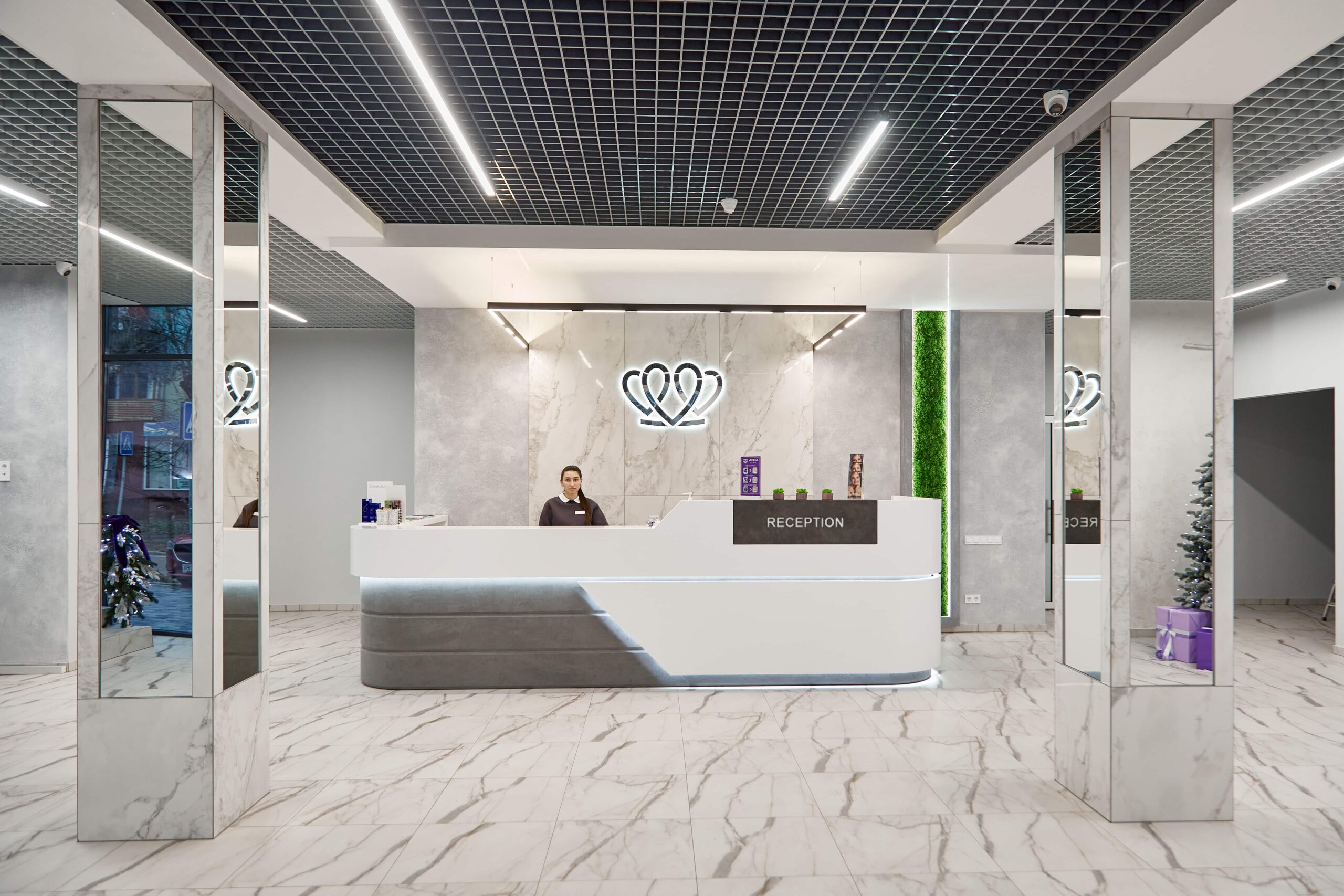
📍 Lviv, Ukraine
A dental clinic in Lviv with a separate doctor for each field: oral surgery and implants, prosthodontics, restorative dentistry, endodontics under a microscope, periodontics, orthodontics and paediatric dentistry.
The only difference is when the implant is placed. With the immediate protocol the implant goes into the socket straight after the tooth is extracted, at the same visit and under the same anaesthetic. With the delayed protocol you wait for the socket to heal: 4–8 weeks for the gum to heal, 12–16 weeks for partial recovery of the bone, or six months or more for full healing. The implant itself, the materials and the prosthetic stage are the same; what differs is the conditions under which the protocol is possible at all and how demanding the extraction technique is.
At the clinics MediUkraine works with, a consultation with a CT scan costs from USD 20, tooth extraction from USD 42, and a Neodent implant with placement from USD 373. Bone grafting, if needed, is a separate line from USD 144. The abutment and the crown are charged separately from the surgical stage. The final total is set by the clinic after an examination and a CT scan, and the preliminary quote is usually drawn up in two versions straight away, for the immediate and the delayed scenario.
The protocol is not used where there is an acute purulent infection with an abscess or fistula, where the buccal socket wall is destroyed, or where there is not enough bone beyond the root tip for the implant to hold straight away. General limitations are uncontrolled diabetes, treatment with bisphosphonates or denosumab, radiotherapy to the jaws, uncorrected blood clotting disorders and jaw growth not yet complete in teenagers. Untreated periodontitis, heavy smoking and poor hygiene are relative: once they are dealt with, implants can be reconsidered.
Because the full picture can be seen only after the tooth is extracted. A scan does not show whether the thin buccal wall will crack as the root comes out, how far the granulation tissue has spread, or what torque can be achieved in that particular bone. If the wall is damaged, the inflammation has reached the bone or the implant does not reach 25–35 N·cm, the surgeon fills the socket with bone material, covers it with a membrane and postpones implant placement for 3–4 months. This switch is built into the plan in advance, and the quote is drawn up for both scenarios from the start.
Sometimes, but it is a separate decision and not for everyone. A temporary crown is fitted on the day of the operation when primary stability is sufficient, roughly from 35 N·cm, and most often in the front area, where preserving the gum contour matters. It is always kept out of the bite: it shapes the gum rather than chewing. The permanent crown is made only after osseointegration, 3–4 months later in the lower jaw and 4–6 months in the upper. With lower stability a healing abutment or a removable temporary denture is used.
Sutures are removed on day 7–10, and the swelling goes down during the first week. Osseointegration takes roughly 3–4 months in the lower jaw and 4–6 months in the upper, longer if a significant volume of bone material was added. The permanent crown is made after a check X-ray and a test of implant stability, not by the calendar. The gum contour stabilises even more slowly: it is finally assessed 6–12 months after the operation.
In terms of implant survival in carefully selected cases the protocols are comparable, but aesthetic complications are statistically more frequent with the immediate option, above all recession of the gum margin in the smile zone. The risk rises with a thin gum biotype and a buccal wall thinner than 1 mm. It is reduced by filling the gap with bone material, thickening the gum with a connective tissue graft and correctly shifting the implant towards the palatal wall. Only a surgeon, after an examination and CT scan, can weigh the risks and benefits in your case.
Tell us what you need: we will suggest a clinic and a doctor and send you a cost estimate.
Request a consultation →