| Service | 🇺🇦 Ukraine | 🇨🇿 Czech Republic | 🇹🇷 Turkey |
|---|---|---|---|
| Professional cleaning under a microscope | from USD 44 | from USD 95 | from USD 99 |
| Complex-case cleaning under a microscope | from USD 59 | — | — |
| Professional cleaning without a microscope | from USD 37 | from USD 72 | from USD 63 |
Prices in the “Ukraine” column come from MediUkraine’s partner clinics. The Czech Republic and Turkey columns show the lower bounds of prices published by clinics there (for Turkey, also the Turkish Dental Association’s recommended 2026 tariff), collected in August 2026 and converted to US dollars at the rate on the day of collection. A dash means we could not find a comparable published price. In any country, the final cost is set by the clinic after an examination.
This is a professional cleaning carried out under a dental operating microscope. The optics give magnification of roughly 3 to 25 times and light directed along the line of sight, so the dentist sees the gum margin, the root surface and the edges of restorations without shadow. The microscope itself removes nothing: the instruments stay the same, but they are used with greater precision.
With the naked eye, the dentist sees roughly what you see in the mirror: plaque, visible tartar above the gums, the colour of the enamel. Magnification reveals the details that decide whether the inflammation will come back: remnants of subgingival tartar at the gum margin, an overhanging edge of an old filling, a gap under a crown margin, an enamel crack, early caries in a fissure, and leftover cement around an implant crown.
Those who benefit most are patients with implants, with a history of periodontitis and with many restorations: fillings, veneers, crowns, bridges. The same goes for people whose tartar comes back within six months, those who wear braces, and those about to have prosthetic work or implants: before these stages, neither inflammation nor a defective margin should be missed.
There is also a flip side, and it is worth saying plainly: not everyone needs magnification. If your gums do not bleed, probing shows pockets of up to 3 mm, you have few or no restorations and you have hygiene appointments twice a year, an ordinary cleaning will give practically the same result. In that case your money is better spent on keeping to regular visits than on optics.
A planned cleaning is postponed if you have an acute respiratory infection, a cold sore flare-up or mouth ulcers. With pockets deeper than 4 mm, a cleaning alone is not enough: separate periodontal treatment is needed, and the microscope does not change that. MediUkraine is a treatment search and coordination service, not a clinic: we choose the dental clinic and the dentist, agree the date of the visit and support you throughout, including if you are travelling from abroad. The scope of treatment is decided by the dentist after an examination.

Travel. Ukrainian airspace is closed, so there are no commercial flights. Patients reach Lviv overland, most often from Poland: Rzeszów and Kraków airports are the usual entry points, with a train, bus or private transfer onward to the border and on to the city. Lviv is roughly 70 km from the Polish border, the shortest crossing of any Ukrainian city.
Visas. Citizens of the EU, the United Kingdom and the United States do not need a visa for short stays in Ukraine. Rules depend on your citizenship, so check your own before booking.
Safety, plainly. Ukraine is at war. Air raid alerts occur in Lviv, and an appointment can be interrupted by one. Several governments advise against travel to Ukraine, and while that advice stands, ordinary travel and medical insurance is often void — confirm your cover with your insurer before you commit to anything.
Language. MediUkraine handles correspondence, scheduling, the written treatment plan and the quote in English. The clinic's team communicates with international patients in English.
Payment. You pay the clinic directly. Our coordination service is free of charge for you.
More on travel, safety and insurance: Safety and travel.
An operating microscope combines two things: magnification and coaxial light. The light travels along the optical axis, so it illuminates the exact point the dentist is looking at and leaves no shadow in a recess or under the gum margin, areas an ordinary dental lamp cannot reach. Magnification is changed in steps: 3–5 times for examination, 6–10 for work, and the highest settings only for brief checks of a particular area, because higher magnification narrows the field of view and reduces the depth of focus.
The main finding under magnification is remnants of subgingival tartar. It is darker than tartar above the gum, firmly attached to the root and lies in the gum pocket, so in an ordinary examination it is detected by touch, as roughness under the probe. Under the microscope, with the gum margin gently retracted, the root surface itself is visible, and the dentist can see whether the area really is clean after ultrasonic scaling. That is the practical difference: not how the tartar is removed, but when the dentist decides that an area is finished.
The second typical finding is an overhanging filling margin on a contact surface. This is a ledge of filling material that extends under the gum and constantly traps plaque: you brush conscientiously, yet the gum in that spot bleeds for years. To the naked eye such a margin is almost invisible; under magnification it is obvious at once. It is smoothed with a strip or a fine-grit bur, as a separate procedure outside the cleaning protocol. A gap under a crown margin and leftover cement around an implant crown (a common cause of inflammation of the surrounding gum) are found the same way.
The third group of findings concerns the hard tissues. Under magnification enamel cracks, early caries in fissures and on contact surfaces, areas of exposed root cementum and wedge-shaped defects at the necks of the teeth become visible. The cleaning does not treat them, but it makes them visible: the dentist records the finding, shows you the image and adds it to the plan. Some, such as a crack without symptoms, are simply monitored and compared at the next visit.
Working under a microscope is more demanding, and this directly affects the length of the appointment. The dentist sees most surfaces in a mirror, so instrument movements are mentally reversed; the field of view is narrow, so each area is covered section by section; and every change of magnification needs refocusing. An assistant holds the saliva ejector and keeps the mirror at the right angle. Altogether the visit takes roughly 20–30 minutes longer than without optics.
The stages themselves are standard and do not depend on the optics. An ultrasonic scaler breaks up the tartar, Gracey curettes finish the subgingival areas, air polishing removes biofilm and staining, then the teeth are polished with paste and fluoride varnish is applied. The microscope comes in at two points: before the work, when the dentist examines each area and plans the scope, and after each stage, to check the result. Around implants and braces a fine glycine or erythritol powder is used, and titanium surfaces are not touched with metal tips.
Now to the limitations, because they often go unmentioned. The microscope cannot see through the gums: if a pocket is deeper than 4–5 mm, its base remains invisible, and such an area is assessed with a periodontal probe, while the state of the bone is assessed on an X-ray. Optics do not treat periodontitis or whiten teeth. Nor do they automatically make the dentist more careful: without mirror skills and an assistant, magnification only slows the appointment down. So whether the clinic has a microscope matters less than whether it is used every day.
So who gains a real benefit from magnification? Patients with implants: leftover cement or plaque at the neck of an implant triggers inflammation of the surrounding gum, which can progress to bone loss and is hard to spot with the naked eye. Patients with a history of periodontitis, for whom every missed remnant of tartar means a pocket coming back. People with many restorations, such as veneers, crowns, bridges and composite fillings on contact surfaces, because plaque collects at the junction between material and tooth. And those facing prosthetic work, implant treatment or orthodontic treatment.
The opposite situation is worth naming honestly too. If your gums do not bleed, probing shows pockets of up to 3 mm, you have few restorations, the plaque is mostly staining, and you have hygiene appointments twice a year, the difference in outcome between the two options will be small. Here it is more useful to keep to regular visits and learn to clean between your teeth than to pay extra for optics. Children and teenagers usually do not need magnification for routine cleanings either.
Finally, frequency: optics do not lengthen the interval between visits. The guideline is the same: every six months for someone with healthy gums and careful home care, and every three to four months with periodontitis, braces, implants, smoking, diabetes or a dry mouth. A clinic cleaning does not replace daily care: the clinic removes the consequences, while home care controls the cause.
Straight after the appointment your teeth feel smooth, and where inflammation was hiding under the tartar the gums may bleed a little. Do not eat or drink for two hours after the fluoride varnish, and do not brush until the next morning, or the varnish will simply come off. For the first few hours it is also best to avoid coffee, tea, wine and smoking: the protective film on the enamel has not yet re-formed, and stains take hold more easily.
Sensitivity to cold and sweet things in the first days is an expected reaction, not a complication. The necks of the teeth and root areas that were covered by tartar for years are now exposed to stimuli for the first time. This usually passes within two to seven days; a toothpaste with potassium nitrate or fluoride, warm water instead of ice-cold, and a break from acidic drinks all help. If sharp pain is concentrated in one tooth or sensitivity lasts more than two weeks, see the dentist.
The gums settle within about a week, and bleeding when brushing should decrease day by day. There is one condition: keep brushing as usual rather than sparing the gums "until they heal". If nothing has changed after two weeks, the cleaning is not the problem: most likely a pocket or a defective restoration margin remains, and you need a follow-up examination.
One sensation alarms many people: after heavy tartar is removed, the teeth look longer and noticeable gaps appear between them. For years the tartar filled the space left by gum recession, and now what was already there has become visible. Slight looseness in the first days has the same cause and usually lessens as the swelling goes down, but it is still worth discussing with the dentist.
The result of the cleaning itself is clear straight away, in the chair: the dentist shows before and after images from the microscope, and they show whether the surface at the gum margin is clean. Anything else the magnification revealed, such as overhanging margins, cracks, early caries or a gap under a crown, goes into a separate plan. Some issues are dealt with the same day and some at later visits, which is normal.
The next visit is usually scheduled six months later, or three to four months later with periodontitis, braces or implants. Comparing images of the same areas at the next visit tells more than any verbal description. If you live abroad, MediUkraine sends you the examination record, periodontal chart and photo record digitally, so that monitoring can continue at home.
Step 1. Examination and periodontal chart. The dentist examines the teeth and gums, runs the probe around each tooth and records the pocket depths and bleeding points, paying attention to overhanging filling margins, crown edges and areas of recession. An X-ray is taken if indicated. At this point the dentist decides whether a cleaning is enough or separate periodontal treatment is needed, and whether pain relief will be required.
Step 2. Microscope examination and photos. The dentist goes area by area at 4–10 times magnification, examining the gum margin, the edges of restorations and the surfaces around crowns and implants, and takes images of what is found. You see the image on screen and immediately understand why a particular spot bleeds. The plan is made at this stage: what is done today and what needs a separate visit.
In total, the visit takes 1 to 1.5 hours, roughly 20–30 minutes longer than a cleaning without optics. Between the initial examination with the periodontal chart and the final fluoride application there are four working stages: ultrasonic scaling breaks up the tartar, curettes under magnification finish the subgingival areas, air polishing removes biofilm and staining, and polishing smooths the surface. After each of them the dentist goes back to the microscope and checks the result, and it is these checks that add time. No hospital stay or recovery period is needed: you can return to your usual activities straight after the appointment, and the only restriction is two hours without food, drink or smoking.
Dental hygiene is not a one-off event but a cycle with two parts: a clinic cleaning at set intervals and daily home care in between. The clinic removes what has already hardened; at home, you stop biofilm from maturing. If only one half works, no optics will help: after a thorough cleaning without home care, tartar comes back within a few months.
If the teeth have been neglected, the first cycle rarely fits into one visit. With heavy deposits, the dentist removes the bulk, gives the gums a week or two to settle, and at the second appointment, under magnification, finishes cleaning what the swelling had hidden. The interval is also needed to see how the gums responded to the first stage.
From there, care either stays at regular cleanings or moves on to periodontal treatment. If, once the deposits are removed, the pockets are no deeper than 3 mm and the bleeding has stopped, regular visits are enough. If there are pockets of 4–5 mm or more, a separate stage of root surface treatment is prescribed, and after six to eight weeks the pockets are measured again and compared with the baseline.
The interval is set by the dentist, and it is not always six months. Three to four months is the usual rhythm for patients with a history of periodontitis, braces or implants, for smokers and for people with diabetes. The rule is simple: the interval is shortened until the dentist finds minimal deposits at each visit.
Cleaning is also often part of another treatment plan. Before implants, prosthetic work, orthodontic treatment and whitening it is the first step, and it is then repeated at set points: before crowns are fitted, after braces are removed, and at maintenance check-ups for patients with implants. These follow-up visits are where magnification is needed most often. MediUkraine plans the schedule so that the dates coincide with your trips.
The most common after-effect is increased tooth sensitivity. Once tartar is removed, areas of the necks and roots that it covered for years are exposed to cold, sweet and sour for the first time. This usually passes within two to seven days, and the fluoride varnish at the end of the visit is there for exactly this reason. Longer-lasting sensitivity occurs with marked gum recession and wedge-shaped defects; in that case the dentist adds a remineralisation course rather than telling you to put up with it.
Bleeding during and after cleaning is not a complication but a sign of inflammation that was already there. Healthy gums do not bleed when touched by an instrument. Working under magnification does carry a risk of its own: when every rough spot is visible, it is tempting to polish the surface to a shine, and excessive instrumentation injures the soft tissue and wears away the root cementum, which is thinner and softer than enamel. Careful work means using curettes where indicated, not just in case.
Air polishing has its own limitations, and magnification helps to respect them. The powder depends on the area: sodium bicarbonate is coarse and is not used on composite fillings, exposed root cementum or implant surfaces; glycine or erythritol powder is used on these areas instead. The jet is not directed at a right angle deep into a pocket or into a fresh wound: rare cases of subcutaneous emphysema have been described, in which air entered the soft tissues and caused sudden swelling of the face. The risk depends on technique, not on the equipment.
Several conditions change the protocol, so mention them before the appointment begins. A pacemaker: most devices are shielded from external interference, but the clinic may plan hand scaling instead of ultrasound. Asthma and chronic lung disease call for more caution with aerosol and water; a salt-free diet means no sodium bicarbonate; clotting disorders and anticoagulants mean a smaller amount of work per visit. A planned cleaning is postponed during an acute infection, a cold sore flare-up or mouth ulcers.
Pregnancy is not a contraindication but a reason not to put things off. Hormonal changes make gum inflammation worse, and that is when deposits do the most harm. A routine cleaning is usually done in the second trimester, when a visit is easier to tolerate; X-rays are taken only when strictly indicated and with a protective apron. During pregnancy a long appointment is broken into shorter stages with breaks, and the chair position is adjusted if the gag reflex is strong.
You have the right to know the limits of the procedure and to get a second opinion. You keep the examination record with pocket depths and the microscope images, and can show them to another specialist: the figures and images are more objective than impressions of the appointment. Contact the dentist without waiting for your next scheduled visit if, after the cleaning, the gum or cheek swells, pain is increasing instead of fading, bleeding lasts more than two weeks or your temperature has risen.
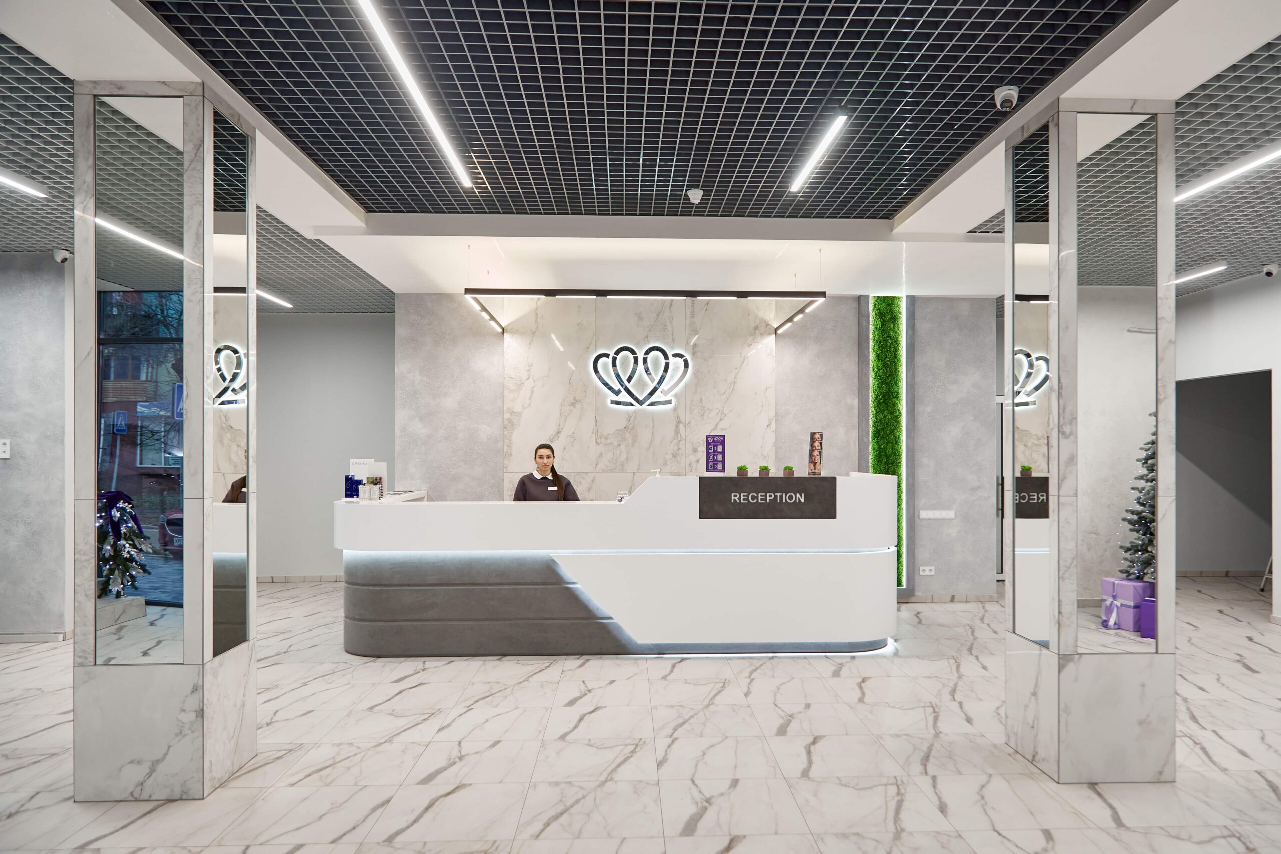
📍 Lviv, Ukraine
A dental clinic in Lviv with a separate doctor for each field: oral surgery and implants, prosthodontics, restorative dentistry, endodontics under a microscope, periodontics, orthodontics and paediatric dentistry.
In terms of stages, not at all: ultrasonic scaling, hand curettes, air polishing, polishing and fluoride application are part of both options. The difference lies in control and time. The dentist examines each area under magnification before starting, returns to the microscope after each stage and checks whether the surface at the gum margin really is clean. These checks make the appointment roughly 20–30 minutes longer. The optics also reveal things that affect the further plan: overhanging filling margins, a gap under a crown, enamel cracks.
At MediUkraine's partner clinic, professional cleaning under a microscope costs from USD 44, complex-case cleaning under a microscope from USD 59, and the same procedure without optics from USD 37. The price includes the dentist's work and the materials for all the stages. Anything beyond the cleaning is charged separately: treatment of caries, X-rays, treatment of pockets deeper than 4 mm, smoothing down overhanging filling margins. Prices are indicative; the dentist confirms the final figure after an examination.
Magnification gives a real benefit to patients with implants, with a history of periodontitis, or with many fillings, veneers and crowns, as well as to people whose tartar comes back within six months and those about to have prosthetic work or implants. If, however, your gums do not bleed, probing shows pockets of up to 3 mm, you have few restorations and you have hygiene appointments twice a year, the difference in outcome will be small; keeping to regular visits matters more. The decision is made by the dentist after an examination and measurement of the pockets.
Usually 1 to 1.5 hours, roughly 20–30 minutes longer than a cleaning without optics, because the dentist stops after each stage to check. With a large amount of deposits the work is split into two appointments a week or two apart. You feel vibration, cold water and pressure; anaesthesia is usually not needed, a numbing gel is applied if your teeth are very sensitive, and occasionally a particular area is numbed. Mention sensitive teeth at the start of the appointment.
First, remnants of subgingival tartar at the gum margin: it is darker than tartar above the gum, and in an ordinary examination it can only be felt with a probe. Next, an overhanging edge of an old filling on a contact surface, which extends under the gum and traps plaque for years, a gap under a crown margin, and leftover cement around an implant crown. Enamel cracks, early caries in fissures and areas of exposed root cementum are also visible. The cleaning does not treat them, but it makes them visible so they can be added to the plan.
No. The optics show the surface, not what lies beneath it: they cannot see through the gums, the base of a pocket deeper than 4–5 mm remains invisible, and the state of the bone can only be seen on an X-ray. That is why pocket depths are measured with a periodontal probe and recorded in a periodontal chart, and an X-ray or CT scan is ordered where indicated. Nor does the microscope treat periodontitis or whiten teeth: cleaning restores the natural colour of the enamel by removing plaque, while changing the shade is a separate procedure.
Yes, in all three cases, with adjustments to the protocol. Around implants and braces, fine glycine or erythritol powders are used, together with tips that do not scratch titanium or ceramics; this is where magnification is needed most often, because leftover cement or plaque at the neck of an implant triggers inflammation of the surrounding gum. The interval between visits for these patients is shortened to three to four months. During pregnancy, a cleaning is usually planned for the second trimester, when a long appointment is easier to tolerate.
Tell us what you need: we will suggest a clinic and a doctor and send you a cost estimate.
Request a consultation →