| Service | 🇺🇦 Ukraine | 🇨🇿 Czech Republic | 🇹🇷 Turkey |
|---|---|---|---|
| Straumann implant | from USD 733 | from USD 929 | from USD 838 |
| Ceramic crown | from USD 280 | from USD 476 | from USD 300 |
| Bone grafting | from USD 144 | from USD 239 | from USD 334 |
| Consultation with CT scan | from USD 20 | from USD 114 | from USD 92 |
Prices in the “Ukraine” column come from MediUkraine’s partner clinics. The Czech Republic and Turkey columns show the lower bounds of prices published by clinics there (for Turkey, also the Turkish Dental Association’s recommended 2026 tariff), collected in August 2026 and converted to US dollars at the rate on the day of collection. A dash means we could not find a comparable published price. In any country, the final cost is set by the clinic after an examination.
The front teeth (the central and lateral incisors and the canines) differ anatomically from the back teeth. The outer bone wall over an incisor root is thin, often less than 1 mm, and gets much of its blood supply from the tooth's own ligament. Once the tooth is extracted, this wall starts to resorb, so the gum contour sinks inwards in the first months after tooth loss, and the longer you wait, the more tissue has to be rebuilt.
Technically, the surgery is the same as for back teeth: a titanium implant is placed in the bone, fuses with it, and a crown is fitted on top. What differs is not the surgery but how the result is judged. For a back tooth it is enough for the implant to be stable and withstand chewing forces; at the front, what also counts is the height of the gum papilla, the symmetry of the gum margin with the neighbouring tooth, the shape and colour of the crown, and whether a grey shadow shows through thin soft tissue.
The procedure is indicated when one or more front teeth have been lost: after an injury, with a vertical root fracture, after failed root canal treatment with chronic inflammation at the root tip, or with a congenitally missing lateral incisor. An implant avoids grinding down the neighbouring teeth for a bridge, which matters here because they are usually healthy teeth that have never been treated.
In some situations the procedure is not done or is postponed. Teenagers and young people whose jaws are still growing do not have implants placed in the front of the mouth: an implant does not move with the jaw, and over time the crown ends up shorter than the neighbouring teeth. Growth ends at roughly 17–18 years in girls and 20–21 in boys, sometimes later, so until then the gap is filled with an adhesive bridge or a removable appliance. Surgery is also postponed in patients with uncontrolled diabetes or untreated periodontitis, during intravenous bisphosphonate or denosumab injection therapy, and after radiotherapy to the jaw area.
MediUkraine is a treatment search and coordination service, not a clinic. We select the clinic and a doctor with the right specialism, agree visit dates and support patients, including those travelling from abroad. Whether this treatment is possible in your case is decided by the doctor after an examination and a review of the CT scan.

Travel. Ukrainian airspace is closed, so there are no commercial flights. Patients reach Lviv overland, most often from Poland: Rzeszów and Kraków airports are the usual entry points, with a train, bus or private transfer onward to the border and on to the city. Lviv is roughly 70 km from the Polish border, the shortest crossing of any Ukrainian city.
Visas. Citizens of the EU, the United Kingdom and the United States do not need a visa for short stays in Ukraine. Rules depend on your citizenship, so check your own before booking.
Safety, plainly. Ukraine is at war. Air raid alerts occur in Lviv, and an appointment can be interrupted by one. Several governments advise against travel to Ukraine, and while that advice stands, ordinary travel and medical insurance is often void — confirm your cover with your insurer before you commit to anything.
Language. MediUkraine handles correspondence, scheduling, the written treatment plan and the quote in English. The clinic's team communicates with international patients in English.
Payment. You pay the clinic directly. Our coordination service is free of charge for you.
More on travel, safety and insurance: Safety and travel.
Depending on timing, there are three protocols. Immediate: the implant is placed in the socket straight after the tooth is extracted, in a single visit. Early: after 4–8 weeks, when the soft tissue has healed but the bone has not yet had time to remodel significantly. Delayed: after 3–6 months, when the socket has healed completely. For front teeth, the choice depends not on convenience but on the condition of the outer bone wall on the CT scan.
Immediate placement shortens treatment and helps preserve the existing gum contour, but the requirements are strict: an intact outer wall of the socket, a thick gum biotype, no acute purulent inflammation and enough bone beyond the root tip for the implant to achieve primary stability. The implant is then placed slightly towards the palate, and the gap between it and the socket wall is filled with slowly resorbing bone graft material. If the wall is damaged, immediate placement is ruled out: the gum margin would recede within a few months, and correcting that would be harder than waiting.
The three-dimensional position of the implant matters more here than anything else. It is standard practice to leave at least 1.5–2 mm of bone on the outer side, about 1.5 mm to the root of the neighbouring tooth and no less than 3 mm between two implants; vertically, the implant is placed about 3 mm below the future gum margin. Placing it a millimetre too far outwards later leads to gum recession and a grey line at the neck, while placing it too deep makes cleaning and soft-tissue support harder. That is why surgeons more often use a surgical guide here rather than working freehand.
The gum biotype, the thickness and shape of the gum margin, determines how predictable the result is. A thin scalloped biotype with tall, pointed papillae is more easily injured and prone to recession; a thick flat biotype is more resilient and more forgiving. It is easy to assess: with a thin biotype, a periodontal probe placed in the gingival sulcus shows through the gum. If the biotype is thin, soft-tissue thickening is planned in advance rather than after recession has occurred.
Grey show-through is a complaint specific to front teeth. The titanium neck of the implant or a titanium abutment casts a cold grey shadow through thin gum tissue at the gum margin, visible when you smile. The risk is reduced in several ways: placing the implant towards the palate, a narrower platform, a zirconia abutment or a hybrid design of zirconia on a titanium base, and above all thickening of the soft tissue with a connective tissue graft from the palate. The graft is taken under local anaesthesia; the donor site heals in about two weeks and is not visible afterwards.
Bone grafting is needed more often at the front than at the back of the mouth, for purely anatomical reasons: after extraction the thin outer wall resorbs and the ridge becomes narrower. A small deficit is corrected at the same time as implant placement, with bone graft material under a membrane, a technique known as contour augmentation. A significant loss of volume is rebuilt as a separate stage 4–6 months before implantation. The material may be a xenograft, an allograft or the patient's own bone. Before surgery the doctor explains the choice and the fact that part of the graft resorbs over time, which is why the volume is slightly overbuilt.
The papilla between the crowns does not always grow back. As a rule of thumb, if the distance from the contact point of the crowns to the bone crest does not exceed 5 mm, the papilla usually fills the space; at 6–7 mm it fills it only partly, and a dark triangle remains between the crowns. The papilla between two neighbouring implants is lower than between an implant and a natural tooth, so two missing incisors in a row are a harder task than one. In that situation it is sometimes better to place one implant and restore both teeth with a cantilever bridge on it.
The soft-tissue profile is shaped after integration. First a healing abutment is fitted: a cylindrical cap that opens a channel through the gum for the future crown. But a round healing abutment creates a round opening, while the neck of an incisor is triangular in cross-section, so for front teeth it is only an intermediate step. Next a temporary crown is made and, over several adjustments, gives the soft tissue the required emergence profile. The temporary crown is kept out of the bite so that it takes no load when the teeth close or the jaw moves sideways. The gum takes 2–3 months to mature, after which its shape is transferred to the laboratory with a custom impression coping: a standard coping would destroy the contour that has already been shaped.
The permanent crown is made of lithium disilicate on a titanium base or of zirconia with a ceramic veneer; both transmit light much as enamel does. Screw retention is preferable: with cement, the excess has to be cleared from under the gum, and residual cement is a common cause of inflammation around implants. Screw retention is only possible when the access channel emerges on the palatal surface, which again depends on implant position: one more reason to plan with a guide.
The last thing to assess before starting is the smile line. If the gum does not show when you smile, a slight asymmetry of the gum margin will go unnoticed. With a high smile line, where the gum is fully visible, the result has to meet a higher standard and more preparation is needed: soft-tissue grafting and longer shaping with a temporary crown are added to the plan more often. Where bone and soft-tissue loss is significant, it is more honest to discuss a screw-retained prosthesis with gum-coloured ceramic or a bridge, which mask the tissue defect where surgical reconstruction is unrealistic. Only a doctor can predict the result in an individual case, after an examination and a review of the CT scan.
For the first two or three days there is swelling, sometimes with bruising on the lip; this is a normal tissue response, not a complication. The doctor prescribes painkillers, cold compresses on the first day, rinses or a gel for the soft tissue and, where indicated, an antibiotic. Eat soft, warm food during this period; do not brush the surgical site, but care for it as the doctor has shown you.
Stitches are removed after 7–10 days, when the doctor also checks how the gum is healing. Avoid putting load on the temporary restoration during this period: do not bite with the front teeth, cut hard food with a knife and eat apples or bread in small pieces. If a graft was taken from the palate, the donor site can be tender for about two weeks, and the doctor often makes a protective plate for that time.
Next comes osseointegration, the fusion of the implant with the bone. In the lower jaw it usually takes 3–4 months, in the upper 4–6, and if bone grafting was done at the same time, the period is longer. Before the prosthetic stage a follow-up X-ray is taken and the stability of the implant is checked; there is no point rushing, because early loading is one of the reasons implants fail to integrate.
Shaping the gum takes another 2–3 months. The temporary crown is adjusted several times during this period, gradually changing the emergence profile, and each time the soft tissue needs weeks to adapt. Patients often feel these visits drag things out, but this is the stage that decides whether the gum margin will match the neighbouring tooth.
After the permanent crown is fitted, the soft tissue still changes: in the first year the gum margin usually recedes by about 1 mm, after which it stabilises. So the final result is assessed at around 12 months, by comparing photographs taken under the same conditions, not by how it feels in the first week.
There is also a long-term effect worth knowing about in advance. Natural teeth slowly continue to erupt throughout life, while an implant stays where it is, so after 10–20 years the crown on it may end up slightly shorter than its neighbours. This is more noticeable in patients who were treated at a young age. This can be corrected by remaking the crown, but the crown should not be thought of as permanent. Check-ups every six months and an annual follow-up X-ray help catch changes early.
Step 1. Consultation and CT scan. The doctor examines the mouth, assesses the smile line, the gum biotype and the shape and colour of the neighbouring teeth, takes clinical photographs and refers you for a cone beam CT scan. The scan shows the thickness of the outer bone wall and whether grafting is needed. You receive a plan with stages, timelines and a quote.
Step 2. Planning and the surgical guide. The CT data is merged with a digital scan of the teeth, the future crowns are positioned virtually, and the implant position is calculated from them: the angle, the depth and where the screw access channel will come out. A surgical guide printed from this plan transfers it to the mouth.
In total, it takes about 6–10 months from the first consultation to the permanent crown. Most of that time is spent waiting rather than in the chair: first for the implant to fuse with the bone, then for the gum to mature around the temporary crown. Because there are only a few treatment days, the work can easily be split into two trips planned in advance.
A front tooth implant is not a single procedure but a cycle spread over months, with long pauses between the active stages. For patients travelling from another city or from abroad, it is convenient to split it into two trips, or three if bone grafting is done as a separate stage.
The first trip is diagnostic and surgical: consultation, CT scan, extraction of the root and augmentation if needed, implant placement, a follow-up X-ray and a temporary restoration. This takes 3–5 days in total, only a few hours of which are spent at the clinic; the rest of the time allows for initial healing and a check-up before you leave.
Then comes the osseointegration period: 3–6 months spent at home. During this time, careful hygiene, avoiding load on the surgical site and contacting the doctor if anything seems wrong are important. A temporary restoration fills the gap, so you do not have to go without a front tooth.
The second trip is prosthetic: uncovering the implant, the healing abutment, the temporary crown and, once the soft tissue has matured, the custom impression coping, try-in and fitting of the permanent crown. Because of gum shaping this stage is often split into two visits 2–3 months apart; for patients who live nearby, it is done in visits every few weeks.
Once the crown is fitted, treatment moves into maintenance: check-ups and professional cleaning every six months, an annual follow-up X-ray and, if needed, advice on improving your hygiene routine. You keep the surgical report, the implant passport and the scans, so follow-up can also be done at home. MediUkraine draws up the travel schedule and stays in touch between trips.
The surgical stage carries the usual risks of an outpatient procedure: swelling, bruising, pain in the first few days and, less often, stitches coming loose or wound inflammation. Failed integration is a separate issue: studies put it at roughly 1–5% of cases. The implant is then removed, the bone is given 2–3 months to heal and, with the patient's consent, the surgery is repeated; the situation is unpleasant but manageable.
At the front of the mouth, aesthetic complications are more noticeable than functional ones. The most common are recession of the soft tissue exposing the edge of the crown, a grey shadow through thin gums, an incomplete papilla leaving a dark triangle between the crowns, and a shade that does not match the neighbouring tooth. Some of these are corrected with soft-tissue grafting or by remaking the crown; others are easier to prevent at the planning stage than to treat later.
Peri-implantitis, inflammation of the tissue around the implant with loss of bone, develops mainly where there is plaque, cement residue under the gum, smoking or untreated periodontitis. It is painless, so it can only be detected at a check-up and on an X-ray. Regular check-ups are not a formality here: bone lost around an implant is difficult and sometimes impossible to restore.
Contraindications may be permanent or temporary. The procedure is not planned while the jaws are still growing, during intravenous bisphosphonates or denosumab injections, after radiotherapy to the jaw area, or with severe uncontrolled diseases of the heart, blood or immune system. It is temporarily postponed with uncontrolled diabetes, untreated periodontitis, acute inflammation in the mouth and during pregnancy. Smoking and severe teeth grinding (bruxism) do not rule out treatment but raise the risk, and the doctor discusses this before starting.
There are anatomical limits too. The nasopalatine canal sometimes occupies exactly the spot where the implant for a central incisor would need to go; with marked loss of bone and soft tissue, restoring the natural gum contour surgically is not always possible. In such cases it is more honest to discuss a bridge or a prosthesis with gum-coloured ceramic than to start treatment that is likely to disappoint.
If pain gets worse after surgery, swelling has not gone down after three days, pus appears or you develop a temperature, contact the clinic without waiting for your next appointment. You have the right to a second opinion: the scans, the surgical report and the implant passport can be shown to another specialist, and if needed MediUkraine can arrange a consultation at another partner clinic.
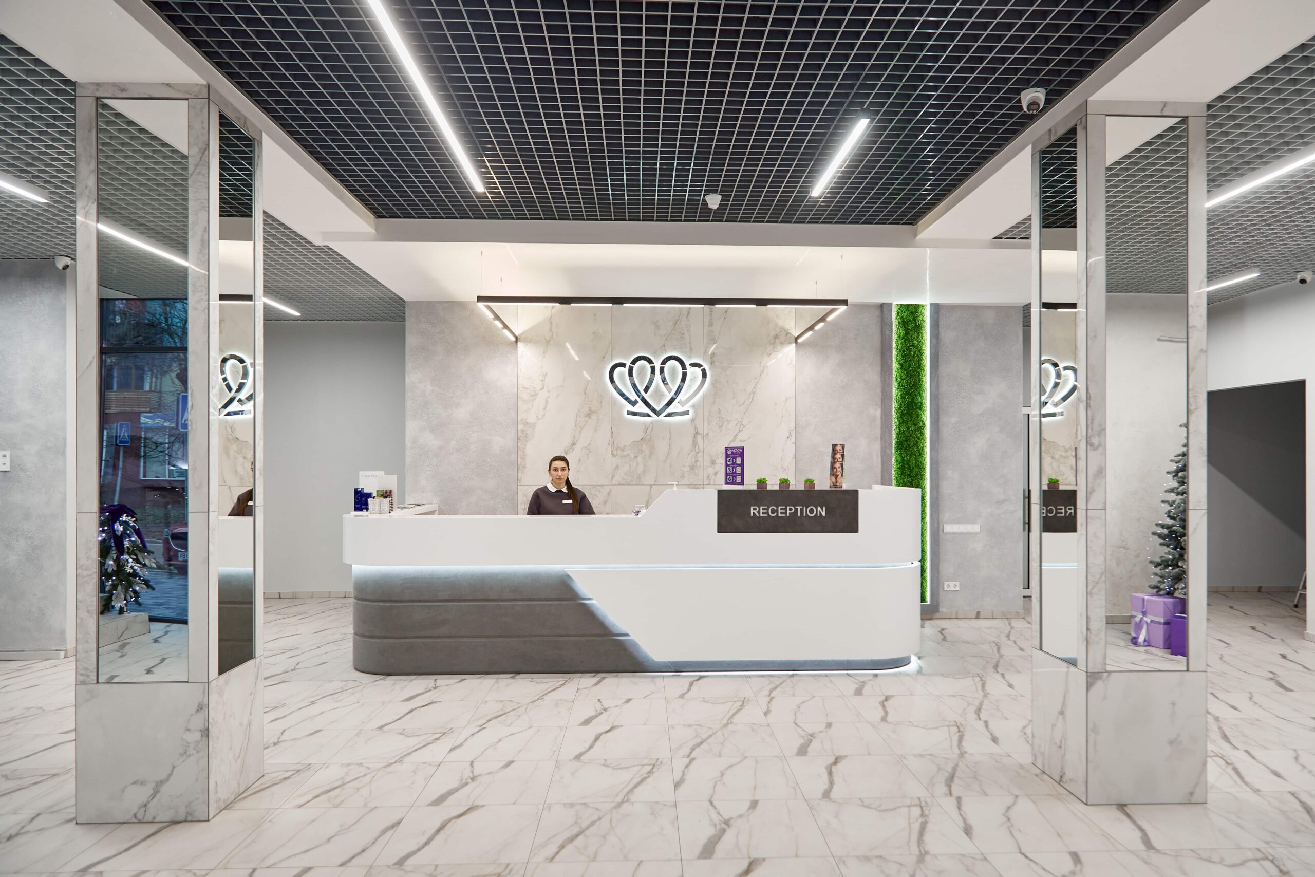
📍 Lviv, Ukraine
A dental clinic in Lviv with a separate doctor for each field: oral surgery and implants, prosthodontics, restorative dentistry, endodontics under a microscope, periodontics, orthodontics and paediatric dentistry.
Sometimes, but the decision is based on the CT scan, not on the patient's wishes. Immediate placement is done when the outer wall of the socket is intact, the gum biotype is thick, there is no acute purulent inflammation and there is enough bone beyond the root tip for the implant to hold stably. Treatment is then shorter and the gum contour is better preserved. If the wall is damaged, the implant is placed after 4–8 weeks or after 3–6 months; otherwise the gum margin will recede, and correcting it would take longer than waiting.
At MediUkraine's partner clinics, a Straumann implant costs from USD 733, a ceramic crown on it from USD 280, bone grafting from USD 144, and a consultation with a cone beam CT scan from USD 20. These are charged separately, so the cost of one tooth includes at least two items, plus grafting, a temporary crown and a surgical guide if needed. The doctor sets the final figure after an examination and a reading of the scan.
In most cases the people you talk to will not notice an implant crown, but the result depends on the starting conditions. It is easier when there is a natural neighbouring tooth to match the shade to, the gums are thick and the smile line is low. It is harder with a thin biotype, significant bone loss and a high smile line, when the gums are fully exposed. The most demanding case is a single central incisor, because the eye instantly compares it with its neighbour. How closely the result will resemble your own tooth is assessed by the doctor at the consultation, from photos and the CT scan.
It is a cold grey shadow along the gum margin: the titanium neck of the implant or a titanium abutment shows through thin soft tissue. It most often appears with a thin gum biotype and with an implant positioned too far outwards. The risk is reduced at the planning stage: the implant is shifted towards the palate, a narrower platform is chosen, a zirconia abutment or a zirconia-on-titanium-base design is used, and the soft tissue is thickened with a connective tissue graft from the palate. If the shadow is already visible, it can be corrected with soft-tissue grafting or by replacing the abutment.
Because of the anatomy. The outer wall of bone over an incisor root is thin, often less than 1 mm, and gets much of its blood supply from the tooth's ligament. After extraction it resorbs, the ridge becomes narrower and the gum margin sinks, and the more time has passed, the more noticeable the difference from the neighbouring area. A small deficit is corrected at the same time as implant placement with bone graft material under a membrane; a significant loss of volume is rebuilt as a separate stage 4–6 months before implantation. The need for grafting is determined from the CT scan.
No, not until the jaws have finished growing. An implant fuses with the bone and does not move with it, while the neighbouring teeth keep moving, so over the years the crown on the implant ends up shorter than its neighbours. Growth ends at roughly 17–18 years in girls and 20–21 in boys, sometimes later, and this is assessed from X-rays and changes over time, not just from the person's age. Until then the gap is filled with an adhesive bridge or a removable appliance, or kept open orthodontically; the decision is made by the doctor after an examination.
Usually 6–10 months. Implant placement takes 40–90 minutes, fusion with the bone 3–4 months in the lower jaw and 4–6 in the upper, longer after bone grafting. Then 2–3 months are spent shaping the gum with a healing abutment and a temporary crown, and only after that is the permanent crown made. There are few active days in the chair, so for patients from other cities treatment is split into two trips, and a temporary restoration fills the gap in the meantime.
Tell us what you need: we will suggest a clinic and a doctor and send you a cost estimate.
Request a consultation →