| Service | 🇺🇦 Ukraine | 🇨🇿 Czech Republic | 🇹🇷 Turkey |
|---|---|---|---|
| Full functional bite diagnostics | from USD 344 | from USD 126 | from USD 150 |
| Gnathologist consultation | from USD 16 | from USD 114 | from USD 110 |
| CT scan, 2 jaws (for clinic patients) | from USD 15 | — | — |
| Treatment plan preparation | from USD 14 | — | — |
Prices in the “Ukraine” column come from MediUkraine’s partner clinics. The Czech Republic and Turkey columns show the lower bounds of prices published by clinics there (for Turkey, also the Turkish Dental Association’s recommended 2026 tariff), collected in August 2026 and converted to US dollars at the rate on the day of collection. A dash means we could not find a comparable published price. In any country, the final cost is set by the clinic after an examination.
The bite is more than the way the teeth meet in one fixed position. The lower jaw moves in three planes: it opens, moves forward and shifts to the side, while the condyles slide along the slopes of the articular eminences. The teeth guide this movement with their inclined surfaces. A routine chairside check shows only where the jaw ends up when it closes, not the path it takes to get there.
A functional examination measures this path for the individual patient and reproduces it outside the mouth. The models of the jaws are mounted in an articulator, which imitates the action of the joints; a face-bow transfers the position of the upper jaw relative to the skull; a centric relation record captures the position of the lower jaw. The dentist can then see premature contacts and interferences that are hard to spot in the mouth.
The examination is recommended before treatment that changes the bite long term: prosthetic work involving six to eight or more units, full-mouth restoration, raising the bite height, implant-supported prosthetics, and orthodontic treatment in adults. The second group of indications is symptoms: pain or clicking in the joint, restricted mouth opening, morning jaw stiffness, pathological tooth wear, bruxism, repeated chipping of ceramics and crowns coming loose.
Sometimes the full protocol is not needed. A single filling, caries treatment, a professional clean and often a single crown on a back tooth can be handled with a routine examination, provided the bite is stable and the joint is healthy. Nor is the examination a treatment in itself: it does not relieve pain, and it replaces neither orthodontic assessment with a cephalogram nor implant planning. If you have acute pain, trismus or a recent jaw injury, that is treated first and the measurements are taken later.
MediUkraine is a treatment search and coordination service, not a clinic. We choose the dental centre and a doctor with the right specialism, arrange the appointment dates and support you throughout, including if you are travelling from abroad. Decisions on the diagnosis and the scope of treatment are made by the doctor after an in-person examination.

Travel. Ukrainian airspace is closed, so there are no commercial flights. Patients reach Lviv overland, most often from Poland: Rzeszów and Kraków airports are the usual entry points, with a train, bus or private transfer onward to the border and on to the city. Lviv is roughly 70 km from the Polish border, the shortest crossing of any Ukrainian city.
Visas. Citizens of the EU, the United Kingdom and the United States do not need a visa for short stays in Ukraine. Rules depend on your citizenship, so check your own before booking.
Safety, plainly. Ukraine is at war. Air raid alerts occur in Lviv, and an appointment can be interrupted by one. Several governments advise against travel to Ukraine, and while that advice stands, ordinary travel and medical insurance is often void — confirm your cover with your insurer before you commit to anything.
Language. MediUkraine handles correspondence, scheduling, the written treatment plan and the quote in English. The clinic's team communicates with international patients in English.
Payment. You pay the clinic directly. Our coordination service is free of charge for you.
More on travel, safety and insurance: Safety and travel.
The face-bow is the first instrument of the protocol. It is a frame positioned using landmarks on the skull: ear pieces sit in the ear canals and a third support rests on a point on the nose or below the eye socket. The bow records the angle at which the upper jaw sits and its distance from the joints, and transfers this relationship to the articulator. Without it the models are mounted arbitrarily, and any error in the tilt of the occlusal plane ends up as a contact that is too high or too low on the finished restoration. A kinematic face-bow, unlike an anatomical one, locates the patient's true hinge axis rather than relying on average points.
Registration of centric relation is the second key stage. Centric relation is the position in which the condyles sit in the upper front part of the joint sockets, and it does not always match the way the teeth habitually close: over the years the muscles have learned to steer the jaw into a comfortable position that avoids interferences. Deprogrammers are used to erase this muscle memory: an anterior JIG, a leaf gauge, a Kois deprogrammer, a bite plate. Some work within minutes, others have to be worn for several days. The resulting position is captured with a silicone or wax bite record.
The articulator imitates the movement of the temporomandibular joints outside the mouth. Average-value instruments work with averaged values for the condylar guidance angle and the Bennett angle, while fully adjustable ones are set individually from the data of an instrumental recording. For a single crown on a back tooth, averaged values are usually enough; for a full-mouth reconstruction they are not, because even a small error is then multiplied across every tooth in the arch. In the instrument the dentist sees premature contacts, interferences on the working and balancing sides, and the pattern of guidance when the jaw moves forward.
Axiography and condylography record the path of movement of the condyle. Mechanical systems draw a graph on a plate, while electronic ones capture the movement with sensors and plot the curve on screen. The recording gives individual angles for programming the articulator and shows any asymmetry between the right and left joints. The method has a clear limit: axiography records movement, not the condition of the tissues, and it cannot replace imaging of the articular disc.
Imaging shows what a clinical examination cannot. Cone beam computed tomography shows the bony structures: the shape and size of the condyle, its position in the socket, erosions, flattening, and the condition of the teeth and the bone around them. MRI is the only method that shows the articular disc itself and whether it is displaced with or without reduction. An orthopantomogram and a cephalogram are added when the plan includes an orthodontic stage.
The clinical examination matters just as much as the instruments. The dentist palpates the chewing and temporal muscles, assesses tenderness at specific points, measures how wide the mouth opens (roughly 40–55 mm is typical in adults), checks whether the jaw moves straight or deviates to the side, listens for clicking and crepitus, and performs joint loading tests. Many clinics work to the structured DC/TMD protocol, which combines a standardised questionnaire with a systematic examination and gives results that can be compared between doctors.
Digital tools complement the classic methods. An intraoral scanner produces models without impression material, a face scan and a photo record link the teeth to the facial features, and a virtual articulator allows the bite to be analysed in software. T-Scan computerised occlusal analysis shows not only where contact occurs but also in what sequence and with what relative force, while electromyography assesses the activity of the chewing muscles. Both are supplementary: a diagnosis is never based on them alone, and the clinic should be upfront about this.
The findings are then turned into a physical plan. In the programmed articulator the technician creates a wax-up of the planned tooth shape, which is transferred into the mouth as a trial mock-up so you can see and assess it before any teeth are prepared. If the plan involves changing the bite height or there is joint dysfunction, a Michigan-type stabilisation splint is made first and usually worn for 6–12 weeks under the dentist's supervision, and only then does treatment move on to temporary restorations.
It is important to be clear about the limits of the method. The examination describes how the bite is arranged, but it does not prove that the bite caused the pain: scientific evidence does not confirm that features of occlusion are the sole cause of temporomandibular joint dysfunction. That is why current clinical guidelines advise starting with reversible measures, a splint, exercises, physiotherapy and adjusting the load, and considering irreversible interventions such as selective grinding or full-mouth prosthetics "to treat the joint" with caution and only for specific indications. Clicking without pain and without restricted movement often needs no treatment at all, only monitoring.
Related treatments usually come after this stage: orthodontic treatment with aligners or braces corrects the position of the teeth, and the prosthetic stage restores their shape with crowns, veneers or implant-supported restorations. The functional examination replaces none of them: it sets the coordinates within which this work is done and the order in which it is done.
You get the dentist's preliminary findings straight after the appointment; the full report takes three to seven working days, because the models have to be cast, mounted in the articulator and analysed, and the imaging reported. That is why the plan is usually presented at a separate visit rather than on the day of the measurements.
If a stabilisation splint is prescribed, it is made within three to seven days. For the first few days your bite will feel unusual, and you may notice extra saliva and slightly unclear speech on some sounds; this usually settles within a few days. The first adjustment is made after 1–2 weeks, and after that the splint is checked once every 4–6 weeks, because the muscles relax and the jaw gradually changes position.
The effect of reversible therapy is usually assessed after at least 6–12 weeks of regular wear. Muscle pain in most cases eases sooner, within 4–8 weeks, whereas clicking caused by a displaced articular disc may persist: the aim of treatment is to remove the pain and restore a normal range of movement, not necessarily to get rid of the sound.
The dentist will also warn you that your bite is expected to change. When the muscles stop holding the jaw in its habitual position, the teeth start to meet differently, and this is not a complication but precisely the aim of this stage. This new position then becomes the basis for prosthetic or orthodontic treatment.
Permanent restorations are almost always preceded by temporary ones, which are used to test the new bite height and tooth shape over 4–12 weeks, and sometimes longer in a full-mouth reconstruction. If anything feels uncomfortable during this time, the temporaries are adjusted: reworking acrylic is much easier than reworking ceramics.
Once the prosthetic work is complete, check-ups are scheduled after one month, after six months and then once a year to check the occlusal contacts, the ceramics and the muscles. Timeframes and progress vary from person to person, so only a dentist can predict the outcome for an individual patient, based on an examination and its findings.
Step 1. Conversation, examination and measurements. The dentist finds out what is bothering you and when it started, what dental work you have already had, and whether there have been jaw injuries or orthodontic treatment. They then examine the teeth for wear and chipping, palpate the chewing and temporal muscles, listen to the joints, and measure how wide the mouth opens and whether the jaw deviates to the side. This part takes up most of the first appointment and determines the scope of everything that follows.
Step 2. Face-bow and registration of centric relation. At the same visit an intraoral scan or impressions of both jaws are taken. The face-bow is then fitted to record the position of the upper jaw relative to the joints, and a deprogrammer is used to remove muscle memory, after which the position of the lower jaw is recorded with a silicone bite record. This is the core of the examination: this data is later transferred to the articulator.
In total the examination fits into two appointments: the first lasts 1.5–2 hours, the second about half an hour. Between them a CT scan of the jaws and joints is taken, the movements of the condyle are recorded with an axiograph where indicated, the models are mounted in the articulator using the face-bow data and the contacts are analysed, and at the second visit the dentist shows the result, explains the findings and hands over a written plan. No anaesthesia, hospital stay or recovery period is needed: straight after the appointment you can eat, talk and return to your usual activities.
The first stage is the examination itself and the plan. It takes two visits over one or two weeks and ends with a document stating what will be done, in what order, how many stages there are and roughly how long each lasts. Without this document there is no point planning the next steps.
The second stage is reversible measures and dental treatment. While you wear a stabilisation splint and follow the advice on jaw loading, any caries is treated, root canals are retreated if needed and gum inflammation is brought under control. Loose or diseased teeth make the bite record unreliable, so this stage cannot be skipped.
The third stage is testing the new position in temporary restorations. The shape and height of the teeth are first reproduced in acrylic for 4–12 weeks, and any corrections are made during this period. If the plan includes an orthodontic stage, it is built in here and takes longer, from a few months to two years depending on complexity.
The fourth stage is the permanent restorations. Before they are made, the parameters are transferred to the articulator again, because the jaw position has changed since the start. Crowns, veneers or implant-supported restorations are tried in, the contacts are checked at rest and in movement, and adjustments are made before the work is fixed permanently.
The fifth stage is maintenance. After extensive work most patients are advised to wear a protective night guard and to have a check-up, including a bite check, every 6–12 months. For international patients the cycle is split into two or three trips, with interim check-ups done remotely from photos and a description of how they feel.
The examination itself is non-invasive: no tissue is cut, no anaesthesia is used and there is no recovery period afterwards. Minor discomfort is possible: a gag reflex when impressions are taken, muscle fatigue from keeping the mouth open for a long time, pressure from the ear pieces of the face-bow. The dentist takes breaks, and if the gag reflex is strong, impressions are replaced with a digital scan.
A CT scan involves exposure to radiation. The effective dose of a cone beam scan of the jaws depends on the machine and the field of view and is usually measured in tens to hundreds of microsieverts, which is comparable to between a few days and several weeks of natural background radiation. The scan is done only when clinically indicated, a protective apron and collar are used, and in pregnant women it is done only when it cannot be avoided, after discussion with the doctor managing the pregnancy.
Deprogrammers and trays require discipline. Anterior bite devices such as the JIG are designed for short-term, supervised use: if such a device is worn round the clock for weeks, the back teeth may lose contact and the front teeth may be pushed into the bone, and instead of helping, the device creates a new bite problem. That is why the dentist sets the wearing schedule and sees you for regular check-ups.
The main risk lies not in the measurements but in the decisions based on them. Irreversible interventions, such as selective grinding of enamel, raising the bite height with crowns or full-mouth prosthetics "to cure the joint", have no reliable evidence base as a way of treating temporomandibular joint dysfunction. If, after the very first examination, you are offered immediate preparation of your teeth, that is a good reason to pause and get a second opinion.
In some situations the examination is postponed or adapted. Acute pain and trismus, a recent injury or a suspected jaw fracture, or a dislocated joint need emergency care first. Active inflammation in the mouth, untreated caries and periodontitis with loose teeth make the bite registration unreliable. Botulinum toxin injections into the chewing muscles in recent months distort muscle tone, so mention them in advance.
The clinics MediUkraine works with hold a licence from the Ministry of Health of Ukraine to practise medicine. You receive a written report and the digital files and are free to seek a second opinion: the scans, models and records can be shown to another specialist. If needed, we arrange a consultation at another partner clinic, and you decide on the scope of treatment yourself, without time pressure.
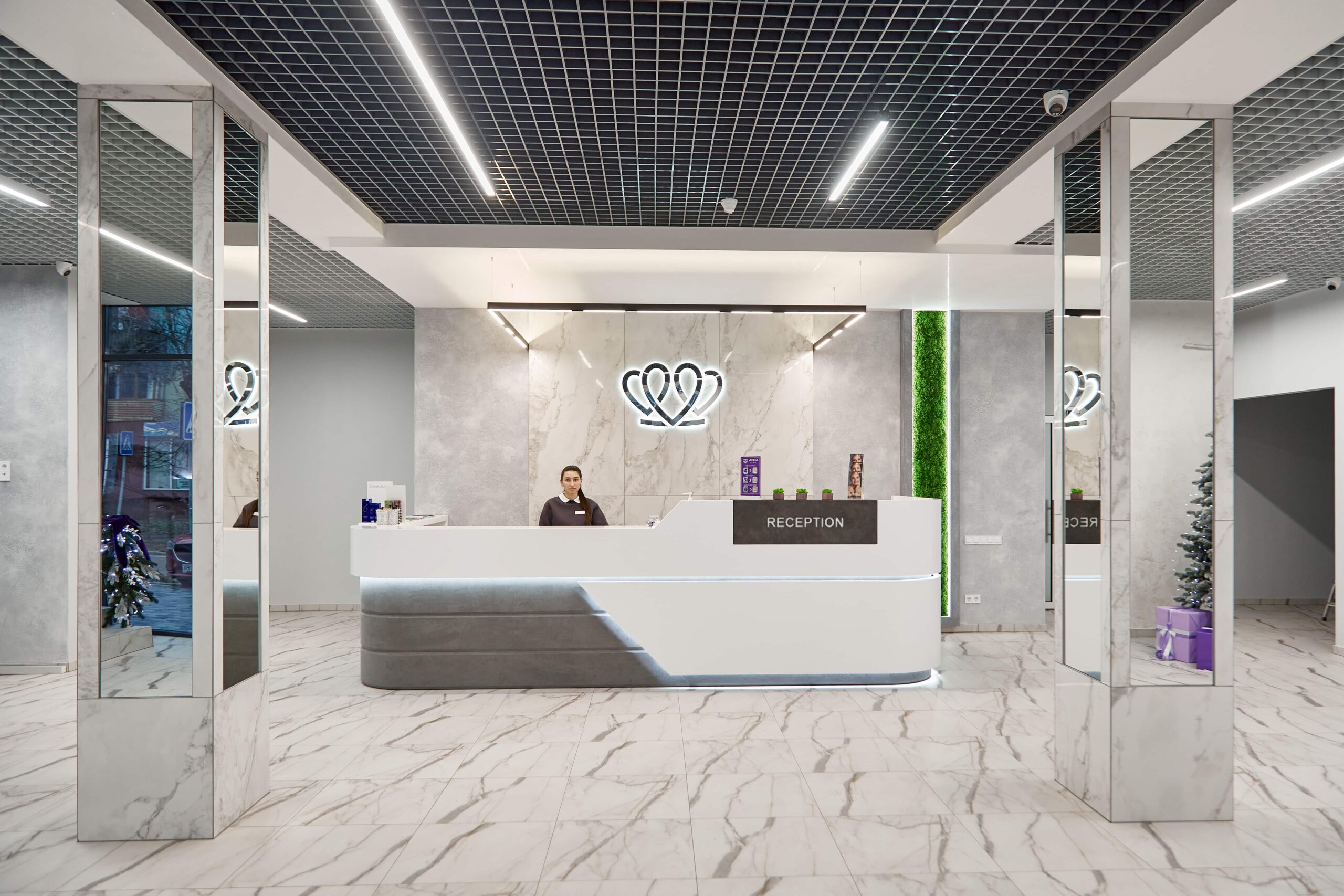
📍 Lviv, Ukraine
A dental clinic in Lviv with a separate doctor for each field: oral surgery and implants, prosthodontics, restorative dentistry, endodontics under a microscope, periodontics, orthodontics and paediatric dentistry.
Crowns and veneers reproduce not only the shape of the teeth but also the path along which the jaw moves. If this path is not measured, the new restoration may keep hitting a premature contact: the ceramic chips, the restoration comes loose, or muscle pain or sensitivity in the supporting teeth develops. The measurements give the dentist the coordinates within which the technician should design the work, and make it possible to check the result on a wax mock-up before the teeth are prepared.
At the clinics MediUkraine works with, the full protocol starts from USD 344 and usually includes a scan or impressions, a face-bow, registration of centric relation, mounting of the models in an articulator and analysis of the contacts. A gnathologist consultation (from USD 16), a CT scan of both jaws (from USD 15) and a written treatment plan (from USD 14) are paid for separately. Splints, wax-ups and any follow-on treatment are charged separately at the clinic's rates. The final cost is confirmed by the clinic.
The examination is painless, and no anaesthesia is needed. What most patients find most unpleasant is the impression material, and that can be replaced with an intraoral scan. The face-bow creates slight pressure around the ears and nose but does not injure the tissues. The full protocol takes 1.5–2 hours with breaks, and the consultation before it 30–40 minutes. After the appointment no recovery or restrictions are needed: you can eat and go back to work straight away.
For a single crown on a back tooth, with a stable bite and healthy joints, the dentist can often manage with ordinary models. But as soon as the work covers several teeth, changes the bite height or involves the front teeth, averaged data is no longer enough: an error in the tilt of the occlusal plane is multiplied with every additional tooth. That is why, before full-mouth restorations, implant-supported prosthetics and adult orthodontic treatment, transferring the measurements to an articulator is considered a standard part of the preparation.
Isolated clicking without pain, without restricted mouth opening and without getting worse over time often needs no treatment: monitoring and keeping the load on the jaw under control are enough. It is worth booking an appointment if pain in the joint or muscles, morning stiffness or headaches in the temples have appeared, or if the mouth has started to open less wide or the jaw locks. Seek help urgently if the mouth suddenly stops opening or closing fully: this may be a disc lock or a dislocation of the joint.
Technically, yes, and for a small amount of work this is normal practice. The problem arises when many teeth are redone at once or the bite height is changed: without measurements the dentist works from averaged values and relies on the patient "getting used to it". If they do not, the finished ceramic work has to be redone, which costs both time and money. Remaking restorations usually costs more than an examination at the outset, which is why it is done before any extensive treatment.
In most cases the pain eases during the reversible therapy stage: with regular splint wear and adjustment of the load, muscle pain usually lessens within 4–8 weeks. At the same time, clicking caused by displacement of the articular disc may remain, and that does not mean failure: the aim of treatment is to relieve the pain and restore a normal range of movement. The course differs from person to person, and only a dentist can predict the outcome for an individual patient, after an examination and analysis of the findings.
Tell us what you need: we will suggest a clinic and a doctor and send you a cost estimate.
Request a consultation →