
| Service | 🇺🇦 Ukraine | 🇨🇿 Czech Republic | 🇹🇷 Turkey |
|---|---|---|---|
| Root canal retreatment | from USD 78 | from USD 234 | from USD 94 |
| Root canal treatment | from USD 67 | from USD 229 | from USD 96 |
| CT scan, 2 jaws (for clinic patients) | from USD 15 | — | — |
| Emergency pain relief | from USD 24 | from USD 72 | from USD 35 |
Prices in the “Ukraine” column come from MediUkraine’s partner clinics. The Czech Republic and Turkey columns show the lower bounds of prices published by clinics there (for Turkey, also the Turkish Dental Association’s recommended 2026 tariff), collected in August 2026 and converted to US dollars at the rate on the day of collection. A dash means we could not find a comparable published price. In any country, the final cost is set by the clinic after an examination.
Retreatment is repeat endodontic treatment of a tooth whose canals have already been filled. The dentist removes the old material from the canals, finds whatever the earlier treatment missed, cleans the canal system again and fills it to its full length. The aim is the same as the first time: to eliminate the space where bacteria live and seal it tightly so that the inflammation at the root tip settles.
Needing retreatment means the earlier work did not achieve its aim, and there are usually several possible reasons. The most common are a missed canal that was left untreated, an underfilled root, where the material did not reach the apical constriction, a ledge or transportation of the canal after working with stiff steel instruments, and a leaking coronal restoration through which saliva has seeped into the filled canals for years.
The indications are a lesion at the root tip that is not shrinking or has appeared after treatment, pain on biting, a fistula on the gum, swelling, an unpleasant taste. Some cases cause no symptoms: the tooth does not bother you, and the changes are spotted by chance on an X-ray before prosthetic work or an implant next to it. Canals are also retreated electively when the tooth is to receive a crown and the old filling material only partly fills the canal.
The method has clear limits. A tooth is not retreated when there is a vertical root fracture: no amount of repeat treatment joins a split root, and the only honest option here is extraction. Nor is treatment undertaken for a tooth broken down below bone level, a tooth that is markedly mobile because of bone loss around it, and sometimes a root whose apical part cannot be reached because of a ledge or hardened paste. In such cases microsurgery is discussed, or extraction followed by prosthetics or an implant.
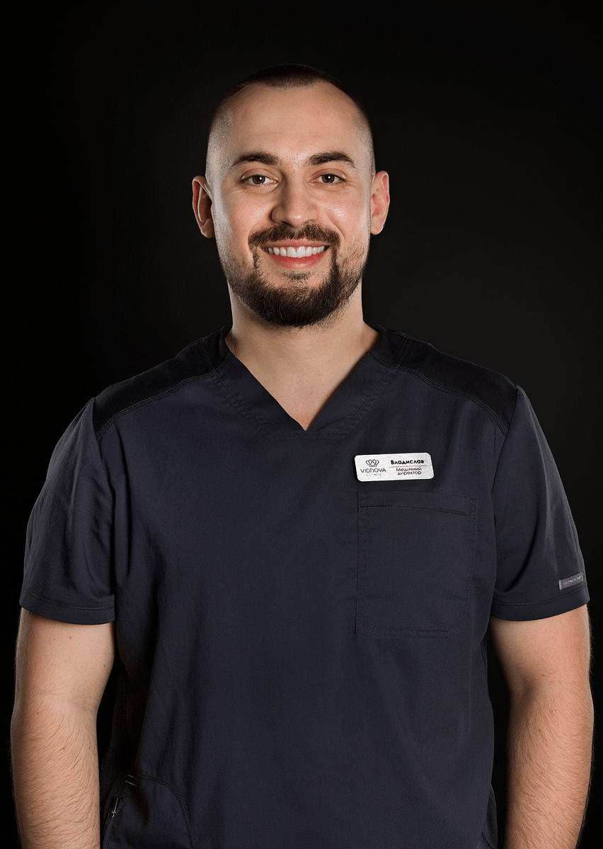
MediUkraine is a treatment search and coordination service, not a clinic. For dental care, we work with our partner clinic, the Vidnova dental practice in Lviv, 10 Ivana-Teodoziia Kurovtsia Street. We select a dentist with the right expertise, arrange the date of the visit and support patients, including those travelling from abroad. The treatment approach is decided by the dentist after an in-person examination and an X-ray.

Travel. Ukrainian airspace is closed, so there are no commercial flights. Patients reach Lviv overland, most often from Poland: Rzeszów and Kraków airports are the usual entry points, with a train, bus or private transfer onward to the border and on to the city. Lviv is roughly 70 km from the Polish border, the shortest crossing of any Ukrainian city.
Visas. Citizens of the EU, the United Kingdom and the United States do not need a visa for short stays in Ukraine. Rules depend on your citizenship, so check your own before booking.
Safety, plainly. Ukraine is at war. Air raid alerts occur in Lviv, and an appointment can be interrupted by one. Several governments advise against travel to Ukraine, and while that advice stands, ordinary travel and medical insurance is often void — confirm your cover with your insurer before you commit to anything.
Language. MediUkraine handles correspondence, scheduling, the written treatment plan and the quote in English. The clinic's team communicates with international patients in English.
Payment. You pay the clinic directly. Our coordination service is free of charge for you.
More on travel, safety and insurance: Safety and travel.
The most common reason for failure is a canal that was never found. The canal system is more complex than the textbook diagram: in upper molars the mesiobuccal root in most cases has a second canal, narrow and hidden under overhanging dentine; in lower incisors a second canal occurs in roughly a third of teeth; and lower molars sometimes have an extra root. An untreated canal remains a reservoir of bacteria: they feed on remaining tissue, exit through the root tip and keep the inflammation in the bone going. From the outside the tooth looks treated, and on a periapical X-ray the filling material appears dense.
The second group of causes is mechanical. The canal may not have been shaped to its full length, stopping a few millimetres short of the apical constriction, or, conversely, material may have been pushed beyond the root tip. A stiff steel file in a curved canal straightens it and creates a ledge beyond which the root tip becomes unreachable; the canal can also be transported to one side, thinning its wall. A filling with voids gives the same result as underfilling: the voids leave a nutrient medium for bacteria.
The third cause lies outside the canal: a leaking coronal restoration. Even perfectly filled canals become reinfected over time if saliva seeps into the tooth for months through a gap under an old filling or a crown whose cement has washed out. So in retreatment the dentist assesses not only the canals but also the condition of the restoration, how well its margins fit and whether there is secondary caries beneath it.
A vertical root fracture is a different matter: retreatment cannot help. It is suspected from a narrow, deep periodontal pocket next to one tooth, a fistula close to the gum margin, pain on biting on a particular cusp, and a characteristic pattern of bone resorption on the CT scan. The fracture is often confirmed only under the microscope once the restoration has been removed, which is why the dentist warns about this possibility in advance: in that case the tooth is extracted rather than treated further.
Retreatment proper begins with dismantling. A crown or a bridge is removed or cut off, a composite restoration is drilled out, a cast post and core is loosened with ultrasound and taken out, and a post is removed with ultrasonic tips, trephines or special extractors. Each of these actions thins the root walls, so the dentist works under magnification and removes only as much tissue as necessary. It is not always possible to save the old crown, and this is worth allowing for in the plan and the quote.
Next comes removal of the old canal filling, and how difficult it is depends on what the tooth was filled with. Gutta-percha is softened with a solvent and withdrawn with retreatment files designed specifically for this purpose, which is the most predictable case. Zinc oxide-based pastes are harder to remove, and the hard materials of past decades, including resorcinol-formaldehyde paste, make the dentine glassy and stain the tooth pink: such canals are negotiated slowly, under the microscope, with ultrasonic tips, and it is not always possible to get through. Cements and fibreglass posts in the canal are drilled out, which adds both time and risk.
An instrument fragment in the canal is a separate task. The fragment is retrieved with ultrasonic tips under the microscope, bypassed from the side and filled into the canal along with it, or left in place if the canal had been cleaned up to the point of blockage. The choice depends on which third of the root the fragment lies in and whether it is beyond a curve: in the straight part of the canal the chances of retrieving it are high, while beyond a sharp curve an attempt often ends in perforation. A fragment in itself is not a death sentence for the tooth: what matters is how clean the canal beyond it has remained.
If the tooth has a perforation, a hole through the wall or floor of the pulp chamber made during earlier treatment or while removing the old filling, it is sealed with a bioceramic material such as MTA (mineral trioxide aggregate). The prognosis depends on the size of the hole, its location and how much time has passed: a fresh perforation in the middle third of the root seals better than an old one at the level of the bone crest, around which inflammation has already developed.
Once the canals have been negotiated to their full length, the main part of the work begins: disinfection. The working length is controlled with an apex locator and confirmed with an X-ray, the canals are shaped with nickel-titanium instruments, and the infection is removed above all by irrigation with sodium hypochlorite activated by ultrasound; EDTA removes the smear layer. In a previously treated tooth there are fewer bacteria, but they are more resistant and hide in the dentinal tubules, so between visits the canals are filled with calcium hydroxide for 7–14 days. Filling is done in the same way as in initial treatment: gutta-percha with sealer to the full length, with no voids, followed by a follow-up X-ray.
The prognosis should be discussed openly: retreatment heals less often than initial treatment. Clinical studies describe healing in roughly 70–80% of cases, compared with 85–90% for initial treatment, and the result is worse where there is a large lesion, a canal that cannot be negotiated, a perforation or little remaining tooth tissue. When retreatment through the crown is no longer possible, microsurgery is discussed, that is, resection of the root tip with a retrograde filling, and with a vertical fracture, destruction below bone level or marked mobility, the more honest solution is extraction followed by an implant. Only the dentist, after an examination and a CT scan, can compare these options for a particular tooth in terms of timeline, cost and likely outcome.
For the first few days the tooth is usually tender on biting: the ligament that holds the root is reacting to the instruments working near the root tip. After retreatment this tenderness can be more marked and last longer than after initial treatment, because old material was moved in the canal and the tissue around the root was disturbed. Discomfort is most noticeable on the first day and eases within 3–7 days; if needed, the dentist recommends an anti-inflammatory painkiller (NSAID) at the usual dose, and it is best not to chew on that side until the permanent restoration is in place.
Some symptoms should not wait for the scheduled check-up: pain that increases instead of easing, swelling of the cheek or gum, a raised temperature, an unpleasant taste from under the temporary filling, or the filling itself falling out. A flare-up after the old filling has been removed happens more often than after initial treatment, and it is usually easy to deal with if you come in the same day rather than a week later.
A temporary filling is designed to last weeks, not months. The permanent restoration of the tooth is planned within 2–4 weeks of filling the canals: it was a leaking coronal restoration that once led to retreatment, so this is not the place to cut costs a second time. A root-filled tooth with little remaining tissue usually needs an onlay or a crown rather than a filling.
The result is judged from an X-ray, not from how it feels: a root-filled tooth does not hurt even while the inflammation at the root tip continues. A lesion in the bone shrinks slowly: the first changes are visible after about 6 months, and the bone takes one to two years to recover, sometimes longer. So follow-up X-rays are taken at 6 and 12 months, and with a large lesion once more after two years, and compared with the CT scan taken before treatment.
The statistics for retreatment are more modest than for initial treatment: studies describe healing in roughly 70–80% of cases. This is a guideline, not a promise: the prognosis for a particular tooth depends on whether all the canals could be negotiated to their full length, on the size of the lesion, on whether there is a perforation and on how much tissue remains. Only the dentist can assess it, after an examination and an X-ray.
If the lesion has not shrunk after a year or the symptoms return, that is no reason to keep waiting. The dentist reviews the situation and proposes the next step: microsurgery with resection of the root tip, or extraction followed by prosthetics or an implant. This scenario is discussed before work begins, so that the patient understands from the outset that retreatment is not the last option for preserving chewing function.
Step 1. Assessment and deciding whether to retreat. The dentist takes the tooth's history, checks the response to biting, probes the gum for a narrow, deep pocket (a sign of a crack) and studies the CT scan. This is where the key questions are answered: how many canals the tooth has and how many of them were filled, whether there is a ledge, a perforation or a fragment, how large the lesion is and whether there is enough tissue for a future restoration. The patient sees the scan, hears about the alternatives (microsurgery or extraction) and receives a quote broken down by stage.
Step 2. Anaesthesia, rubber dam and dismantling the old restoration. The tooth is anaesthetised and isolated with a rubber dam, after which the crown is removed or the filling drilled out, secondary caries is cleared and the remaining tissue is assessed. It is at this stage, under the microscope, that a crack the CT scan did not show may become visible, and the plan may change right there in the chair; the patient is warned about this scenario before starting.
In total, a treatment visit lasts 1.5 to 3 hours, and there are usually two or three appointments. Between anaesthesia with dismantling of the restoration and the final X-ray lies the main work: removing the post or the cast post and core, clearing the canals of old gutta-percha or hardened paste, finding the missed canal under the microscope, shaping with nickel-titanium instruments with sodium hypochlorite irrigation and a temporary calcium hydroxide dressing. No hospital stay is needed, you can return to your usual activities the same day, and the permanent restoration is planned as a separate visit.
The cycle starts with a CT scan and ends not with filling the canals but with a follow-up X-ray a year later. Between the first appointment and a fully restored tooth there are usually three weeks to two months, depending on how many visits the canals need and which restoration has been chosen.
The typical scenario is two or three appointments. At the first, the restoration is removed, the post is taken out, the old canal filling is removed, the canals are shaped and calcium hydroxide is left under a temporary filling. After 7–14 days the canals are filled permanently and a follow-up X-ray is taken. A third visit is needed if the symptoms have not settled or a canal could not be negotiated the first time.
The prosthetic stage is a separate part of the cycle that should not be put off. An onlay or a crown is planned within 2–4 weeks of filling the canals: a temporary filling is not designed to last longer, and a leaking restoration is one of the reasons the tooth needed retreatment in the first place.
Some of the work can be done before you travel. The consultation is held remotely using existing X-rays, the plan and quote are agreed in advance, and only the clinical stages remain for the trip, with a known list and duration. A patient from abroad arrives for specific procedures, not to find out what is needed.
After that the cycle moves into monitoring: an X-ray at 6 and 12 months, with a large lesion once more after two years, plus routine hygiene every six months with a check of the restoration. A root-filled tooth does not warn you with pain, so it is the X-ray that shows whether the lesion is shrinking. If there is no progress, the dentist discusses microsurgery or extraction rather than simply continuing to monitor.
A perforation is the most common risk specific to retreatment. A hole through the wall or floor of the pulp chamber occurs while searching for a canal opening under an old restoration, while removing a post or while negotiating a canal with a ledge. It is sealed with a bioceramic material such as MTA, and the sooner after it occurs, the better the prognosis. Magnification and a CT scan before a complex case reduce the risk, but cannot eliminate it.
Instrument fracture in a previously treated canal happens more often than in an untreated one: files and ultrasonic tips work in a narrow space, often beyond a curve and next to old material. What happens next depends on the stage of treatment: the fragment is retrieved with ultrasound under the microscope, bypassed, or left in place and monitored. What matters is that the patient is told and it is recorded in the treatment notes, rather than the dentist carrying on in silence.
A separate risk of retreatment is thinning of the root walls. Every removal of an old filling, widening of a canal opening and removal of a post takes away dentine, and a root with thin walls withstands chewing load less well and is prone to a vertical fracture. Sometimes the root cracks during the removal of a cast post and core itself, and then work is stopped and the tooth is extracted. For this reason the dentist plans the restoration so that the load on such a tooth is reduced.
Rarer but documented complications are extrusion of filling material beyond the root tip and leakage of irrigating solution into the surrounding tissue. The latter causes sharp pain and swelling that pass within a few weeks under the dentist's supervision; that is why the concentration of the solution, the pressure on the syringe and the isolation are not treated as minor details. Temporary numbness of the lip after work on lower teeth may be linked to the root tip lying close to the inferior alveolar nerve.
The main limitation of the method should be understood before starting: retreatment does not always save the tooth. A canal may turn out to be impassable, a crack may become apparent only after the crown is removed, and a lesion may not shrink despite technically correct work. Pain that lasts more than two weeks or returns months later is investigated with an X-ray, not by trying different painkillers. That is why microsurgery and extraction are discussed as part of the plan from the outset, not as a surprise at the end.
Contraindications mostly concern not the procedure itself but the condition of the tooth and the patient. Work is not started with a vertical root fracture, destruction below bone level or marked mobility of the tooth because of bone loss around it. Pregnancy, clotting disorders, taking anticoagulants or bisphosphonates, and allergies to anaesthetics and latex should be mentioned to the dentist before starting: planned stages during pregnancy are usually carried out in the second trimester, and X-rays are taken with a protective apron. The patient has the right to a second opinion: the CT scan, the treatment record and the quote stay with the patient, and they can be shown to another specialist before the old restoration is removed.

📍 Lviv, Ukraine
A dental clinic in Lviv with a separate doctor for each field: oral surgery and implants, prosthodontics, restorative dentistry, endodontics under a microscope, periodontics, orthodontics and paediatric dentistry.
Before the treatment itself, the previous work has to be dismantled: the crown or filling removed, the post or cast post and core taken out, the old gutta-percha dissolved or the hardened paste negotiated, a ledge bypassed, a missed canal found, and sometimes an instrument fragment retrieved. Each step takes time and carries its own risk, and there is less dentine left in the root than there was at the start. So retreatment takes longer, costs more and, statistically, has a lower healing rate.
At MediUkraine's partner clinic, retreatment of one root canal costs from USD 78, initial treatment of a canal from USD 67, a CT scan of both jaws from USD 15 and emergency pain relief from USD 24. The price is calculated per canal, so work on a molar with three or four canals costs more than work on an incisor. Removing a crown, taking out a post and restoring the tooth are paid for separately. The prices are indicative; the final figure is set by the dentist after an examination and a CT scan.
Most often the reason is that infection has remained in the tooth: a canal that was not found last time, a part of the root the filling material did not reach, or saliva that has seeped under a leaking crown for years. Less often the cause is a vertical root fracture or excessive load on the tooth from its restoration. A root-filled tooth may not hurt at all while the inflammation at the root tip continues, so the cause is established from an X-ray, not from how it feels.
No. Retreatment is not done if there is a vertical root fracture, if the tooth is broken down below bone level, or if it is markedly mobile because of bone loss around it. A technical limit can be a canal that cannot be negotiated: hardened paste, a sharp curve beyond a ledge, or an instrument fragment that can neither be retrieved nor bypassed. In such cases the dentist discusses microsurgery with resection of the root tip, or extraction followed by prosthetics or an implant.
Usually two or three. At the first, the old restoration and post are removed, the old canal filling is taken out and the canals are shaped, after which calcium hydroxide is left under a temporary filling. After 7–14 days the canals are filled permanently and a follow-up X-ray is taken. A third appointment is needed when the symptoms have not settled or a canal could not be negotiated the first time. A treatment visit lasts 1.5 to 3 hours, and the permanent restoration is planned separately, within 2–4 weeks.
In most cases, yes: without removing it the dentist cannot see the floor of the pulp chamber, find a missed canal or assess the tissue under the restoration. Sometimes the dentist works through the crown and keeps it, for example with crowns on front teeth without a post, but the access hole then has to be sealed, and such a crown seals less well. The decision is made by the dentist based on the CT scan and the condition of the restoration; the possibility of keeping the crown is worth discussing before work begins.
A fragment in itself is not a death sentence for the tooth: what matters is how clean the canal beyond it has remained and whether there is inflammation at the root tip. A fragment in the straight part of the canal is usually retrieved with ultrasonic tips under the microscope; beyond a sharp curve it is more often bypassed from the side and the canal filled along with it. If it can neither be retrieved nor bypassed, resection of the root tip or extraction is discussed. Trying to retrieve a fragment at any cost carries a risk of perforation and root fracture.
Tell us what you need: we will suggest a clinic and a doctor and send you a cost estimate.
Request a consultation →