| Service | 🇺🇦 Ukraine | 🇨🇿 Czech Republic | 🇹🇷 Turkey |
|---|---|---|---|
| CT scan, 2 jaws (for clinic patients) | from USD 15 | — | — |
| Panoramic X-ray | from USD 9 | from USD 23 | from USD 15 |
| Consultation with CT scan | from USD 20 | from USD 114 | from USD 92 |
| Treatment plan | from USD 14 | — | — |
Prices in the “Ukraine” column come from MediUkraine’s partner clinics. The Czech Republic and Turkey columns show the lower bounds of prices published by clinics there (for Turkey, also the Turkish Dental Association’s recommended 2026 tariff), collected in August 2026 and converted to US dollars at the rate on the day of collection. A dash means we could not find a comparable published price. In any country, the final cost is set by the clinic after an examination.
Cone beam computed tomography (CBCT) is an X-ray examination in which the X-ray source and a flat detector make one rotation around the head in 10–30 seconds. The scanner takes several hundred projections, and the software builds them into a three-dimensional model of the jaws, which can then be viewed in any plane and measured.
This is what sets it apart from ordinary X-rays. Periapical and panoramic X-rays give a flat image in which the roots, the bone and neighbouring structures overlap one another, and sizes are distorted by magnification. On a CBCT scan the doctor views the area in three mutually perpendicular planes, measures the height and thickness of the bone in millimetres and sees what on a flat image is hidden behind another shadow.
The indications are specific: planning implants and bone grafting, assessing the floor of the maxillary sinus before a sinus lift, impacted and malpositioned wisdom teeth and canines, root canal retreatment, a suspected cyst or granuloma, root resorption, jaw trauma, a suspected root fracture, complex orthodontic cases.
It is just as important to understand when the examination is not needed. Detecting caries, assessing the fit of a filling or checking the bone level around a single implant is cheaper and more accurate with a periapical X-ray. A CBCT scan is not done "just in case" or "for the full picture": there must be a specific clinical question. It shows soft tissue, the salivary glands, the joint disc and tumours outside the bone poorly; these need ultrasound or MRI.
MediUkraine is a treatment search and coordination service, not a clinic. We select a clinic with the right equipment, such as the Vidnova dental practice in Lviv at 10 Ivana-Teodoziia Kurovtsia Street, arrange the date and support patients, including those travelling from abroad. Whether the scan is needed, and its scope, is decided by the doctor after an examination.

Travel. Ukrainian airspace is closed, so there are no commercial flights. Patients reach Lviv overland, most often from Poland: Rzeszów and Kraków airports are the usual entry points, with a train, bus or private transfer onward to the border and on to the city. Lviv is roughly 70 km from the Polish border, the shortest crossing of any Ukrainian city.
Visas. Citizens of the EU, the United Kingdom and the United States do not need a visa for short stays in Ukraine. Rules depend on your citizenship, so check your own before booking.
Safety, plainly. Ukraine is at war. Air raid alerts occur in Lviv, and an appointment can be interrupted by one. Several governments advise against travel to Ukraine, and while that advice stands, ordinary travel and medical insurance is often void — confirm your cover with your insurer before you commit to anything.
Language. MediUkraine handles correspondence, scheduling, the written treatment plan and the quote in English. The clinic's team communicates with international patients in English.
Payment. You pay the clinic directly. Our coordination service is free of charge for you.
More on travel, safety and insurance: Safety and travel.
The field of view (FOV) is the first thing the doctor chooses before the scan, and it determines both the amount of information and the dose. A small field of 4×4 or 5×5 cm covers a few neighbouring teeth and is used in endodontics, for an impacted tooth, and for suspected resorption or a root fracture. A medium field of 8×8 or 10×10 cm covers one jaw, a large field of 13×15 cm covers both jaws together with the sinuses, and an extended field also takes in the temporomandibular joints. The rule is simple: the field is chosen for the question, not "just in case".
The second parameter is voxel size, that is, the edge length of a volume pixel. Modes with a 75–100 µm voxel are needed where fine detail matters: an additional canal, a fracture line, a narrow periodontal space. For implant planning 200–300 µm is enough, and such a mode means a shorter scan and a lower dose. A finer voxel does not automatically make the image better: it increases noise and requires the patient to keep even stiller.
The most common reason for the scan is bone volume. A panoramic X-ray shows only the height of the ridge, and even that is distorted by magnification, while the thickness is not visible at all. A CBCT scan gives both figures in millimetres on a cross-section exactly at the point of the future implant: how much bone height is left to the anatomical boundary, whether the width is enough for the chosen diameter, what the slope of the ridge is, whether there is a concavity on the tongue side. This is where it is decided whether bone grafting is needed and whether it can be done at the same time as implant placement.
The inferior alveolar canal is another reason a CBCT scan is needed. It carries the nerve responsible for sensation in half of the lower lip and chin, and its position varies from person to person: the canal may lie closer to the tongue-side or the cheek-side wall, sometimes splits in two, and in front of the mental foramen often forms an anterior loop that runs a few millimetres forward. This loop is not visible on a flat image, and missing it is what leads to nerve injuries during implant placement in the side of the jaw. The distance from the canal to the root of a wisdom tooth is measured just as precisely before extraction.
Additional roots and canals are a typical finding in endodontic retreatment. A second mesiobuccal canal in upper molars occurs in more than half of teeth, lower incisors may have a second canal, and some lower molars have an additional lingual root. If such a canal has been left untreated, the tooth keeps causing trouble after seemingly completed treatment, and it is the CBCT scan that shows why. It also shows the curvature and length of the root and the relationship of a lesion to neighbouring structures.
Cysts, granulomas and resorption can only be assessed in three dimensions. A periapical X-ray shows a lesion mainly once the cortical plate has already been destroyed, so small changes near the buccal roots are often not visible on it at all. The slices show the true size of a lesion in three dimensions, its boundaries, its relationship to the canal and the sinus, and the depth of external cervical resorption, a condition in which the decision to keep or extract the tooth depends on how close the defect has come to the pulp.
Before a sinus lift the maxillary sinus is examined. Three things matter: the height of the patient's own bone under the sinus floor, the presence of bony septa inside it, which raise the risk of tearing the membrane during the lift, and the condition of the sinus lining itself. Marked thickening of the lining or a blocked natural opening of the sinus changes the sequence: first a consultation with an ENT specialist, and only then bone grafting.
Now for the dose, without pretending it is zero. The effective dose for a small-field scan is roughly 20–70 µSv, and for a scan of both jaws roughly 70–200 µSv depending on the scanner and the mode. For reference: a digital periapical X-ray is a few microsieverts, a panoramic one about 15–25 µSv. Natural background radiation, which everyone receives simply from the soil, building materials and cosmic radiation, averages about 2.4 mSv a year, that is roughly 6–7 µSv a day. So a small-field scan roughly equals a few days of normal background exposure, and a scan of both jaws roughly ten days to a month. The amount is small, but not zero, which is why the scan is done only when indicated, not to complete the records.
The method has clear limits. Metal produces streak artefacts: posts, cast cores, amalgam and bulky crowns spoil the slices around them, so a CBCT scan cannot always confirm a root fracture in a tooth with a post. The grey values in the image are not true Hounsfield units, as in medical CT, so bone density can be assessed from them only approximately. Soft tissue, the joint disc and the salivary glands need other examinations. And any movement while the scanner rotates blurs the images so the scan has to be retaken.
The patient receives the result in DICOM format, on a disc, a USB stick or via a link, together with viewer software. This really matters: DICOM contains all the slices and allows any subsequent doctor to take their own measurements, whereas printed images or JPG screenshots are no good for planning. A written report from a radiologist covering all the areas within the field of view is added to the files. The same file, combined with an intraoral scan, is used to make a surgical guide for implant placement.
There are no restrictions immediately after the examination. No anaesthesia was used and no contrast was given, so you can drive, go to work or go on to the next procedure straight away. You feel nothing, either during the scan or afterwards.
The initial reconstruction takes a few minutes: the software assembles the volume and builds a panoramic view and cross-sectional and axial slices. A detailed written report from a radiologist is usually ready within half an hour to two working days, depending on workload and the size of the field of view. If the scan is taken on the day of a consultation, the dentist looks at the images straight away, without waiting for the formal report.
The scan is then reviewed with the treating doctor. It is not a good idea to interpret the slices yourself: an area of reduced density in the bone may be a lesion or an anatomical variant, and thickening of the sinus lining by a few millimetres occurs in a significant proportion of people without any symptoms. The doctor decides what it means by comparing the scan with the examination and your symptoms.
How long a scan stays valid depends on what has happened to the jaw. For implant planning a scan is usually considered current for up to 6 months. But if a tooth has been extracted, bone grafting done or a sinus lift performed in that time, the earlier scan no longer reflects the actual bone: after an extraction the ridge loses some of its width during the first months of healing, and planning cannot be based on the old figures.
Follow-up after implant placement usually does not need a repeat CBCT scan. The bone level around a placed implant is checked with a periapical X-ray, and it is this that shows changes over the years. A repeat CT scan is ordered for a new question: a suspected complication, planning of the next stage, a clinical picture that does not match expectations.
It is worth keeping the file yourself, not only on the storage device you were given but also as a copy in the cloud. You will need it when seeing another specialist, for a second opinion and when treatment continues in another city or country. MediUkraine passes the files and the report on to you exactly as the clinic issued them, without converting them into images.
Step 1. Examination and the question for the scan. The doctor examines the mouth, looks at existing X-rays and defines exactly what the scan should show: whether there is enough bone for an implant in a specific position, how many canals a tooth has, how the nerve runs relative to the root of a wisdom tooth. Without a clear question the scan becomes aimless screening, and the patient still receives the radiation.
Step 2. Choosing the field of view and the mode. For that question the radiographer sets the field size, the voxel size and the exposure parameters, adjusted for the patient's age and build. For a child the field is limited more strictly than for an adult. This is the most important step as far as dose is concerned: a properly limited field can reduce the exposure several times over compared with a default scan of both jaws.
In total the visit takes about 15–20 minutes: between posing the clinical question and reviewing the finished scan come choosing the field of view for that question, removing metal objects and positioning the head, the scan itself lasting 10–30 seconds, and computer reconstruction with a check for motion artefacts. No anaesthesia, hospital admission or recovery period is needed: you can return to your usual activities straight after the scan.
A CBCT scan is rarely a stand-alone service: it is a stage within longer treatment. So from the outset it is worth understanding how many times, and at which points, you will need to return to the X-ray room.
The first scan is done at the planning stage, before any procedures. It is the basis for decisions on the number and positions of implants, the need for bone grafting, whether to keep doubtful teeth and whether root canal retreatment makes sense. If the plan involves a surgical guide, this same scan is sent to the laboratory together with an intraoral scan.
A second scan is needed when the bone has changed. After a sinus lift or bone grafting a check is usually scheduled after 4–6 months, once the material has remodelled, and it is on that scan that it is decided whether an implant can be placed and of what size. A new scan is also taken after a tooth has been extracted with a view to placing an implant later.
Between these points, follow-up mostly uses two-dimensional X-rays. The progress of root canal treatment, the seating of an implant and the fit of a crown are checked with periapical X-rays: they are quicker, cheaper and give a dose tens of times lower. A CBCT scan is repeated only for a new question, not on a schedule.
Overall, over a full implant cycle from planning to crown, a patient usually has one or two CBCT scans and several periapical X-rays. Exactly how many depends on the scope of work and whether bone grafting is needed. MediUkraine builds these visits into the schedule in advance, so patients from abroad know how many trips they need before the first one.
The main, and essentially the only, risk of the scan is ionising radiation. The dose is small but not zero, and it accumulates, so diagnostic radiology follows the principle of justification: a scan is taken when its result can change the treatment approach. The practical rule is "as low as diagnostically acceptable, taking into account the indication and the individual patient": the field of view and the mode are chosen for the question rather than set to maximum.
Children and teenagers are more sensitive to radiation than adults, because their tissues are growing and they have more years of life ahead. This is not a ban: for an impacted canine, a supernumerary tooth, trauma or a complex orthodontic case the examination is justified. But a child's field of view is limited more strictly, the mode is reduced, and routine CBCT scanning before ordinary orthodontic treatment without specific indications is not done.
Pregnancy is a relative contraindication. An elective scan is postponed, and an urgent one is performed on the doctor's decision with the field limited as far as possible: the jaws are far from the uterus, and scattered radiation barely reaches the foetus, but unnecessary exposure during pregnancy is avoided on principle. Breastfeeding creates no restrictions at all: contrast agents and radiopharmaceuticals are not used here.
There are no absolute contraindications in the usual sense. The only real obstacle is being unable to keep still for 10–30 seconds: a marked tremor, severe involuntary movements, the period after a stroke, severe dementia, or a child under three or four years old. People with claustrophobia usually tolerate the examination calmly, because the scanner is open. Wheelchair users should check in advance whether the clinic has a scanner that works in a seated position.
Another risk lies not in the procedure itself but in misreading the scan. Metal artefacts mimic a root fracture, noise at a fine voxel size looks like changes in the bone, and the grey values on a cone beam image are not true density units, so a conclusion of "soft bone" based on them alone is incorrect. That is why the scan must be read by an appropriately trained doctor, and uncertain findings checked against the clinical picture and symptoms.
A large field of view also brings incidental findings. The field takes in the sinuses, the nasal passages, the area of the carotid arteries and the cervical vertebrae, and thickening of the lining, calcifications or other changes that do not bother the patient are often visible there. Some of them need only monitoring, others a consultation with an ENT specialist or a vascular surgeon. That is why the report must be in writing and cover the whole field, not just given verbally: you then have a document you can take to another specialist for a second opinion.
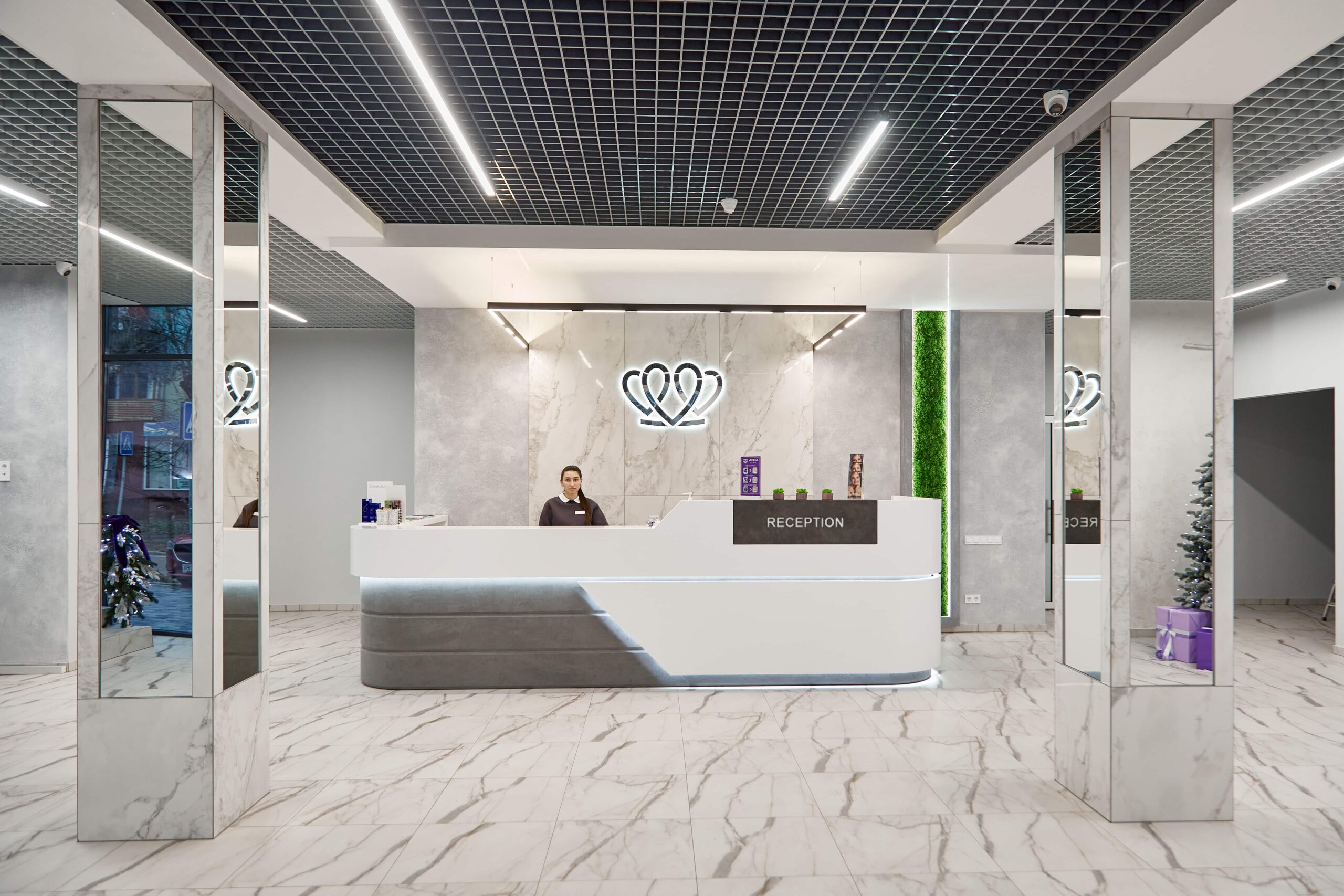
📍 Lviv, Ukraine
A dental clinic in Lviv with a separate doctor for each field: oral surgery and implants, prosthodontics, restorative dentistry, endodontics under a microscope, periodontics, orthodontics and paediatric dentistry.
A panoramic X-ray gives one flat unrolled view in which structures overlap one another and sizes are distorted by magnification. A CBCT scan shows bone thickness and the slope of the ridge in millimetres, the exact position of the inferior alveolar canal and its anterior loop, additional roots and canals, the true size of a lesion or cyst in three dimensions, and bony septa and the condition of the lining in the maxillary sinus. This is exactly the information needed for planning implants, a sinus lift, root canal retreatment and removal of impacted teeth.
At MediUkraine's partner clinics, a scan of both jaws costs from USD 15 and a panoramic X-ray from USD 9. A consultation to review the scan is charged separately, from USD 20 and drawing up a treatment plan from USD 14. The total depends on the size of the field of view, the scan mode and whether a written report from a radiologist is included in the price. A copy of the files on a storage device is usually included in the price of the scan, while printing slices is charged separately. The final cost is set by the clinic after an examination.
The effective dose depends on the field of view and the mode. Scanning a small area of a few teeth gives roughly 20–70 µSv, and a scan of both jaws roughly 70–200 µSv. For reference: a digital periapical X-ray is a few microsieverts, a panoramic one about 15–25 µSv. Natural background radiation from the soil, building materials and cosmic radiation averages about 2.4 mSv a year, that is 6–7 µSv a day. So a scan of both jaws roughly corresponds to ten days to a month of ordinary background radiation.
There is no fixed limit such as "so many times a year": each scan must be justified on its own merits. The question is not how many scans you have had but whether the result can change the treatment approach. Over a full implant cycle there are usually one or two CBCT scans: the first at the planning stage, the second 4–6 months after bone grafting or a sinus lift. Interim follow-up is done with periapical X-rays, whose dose is tens of times lower.
Children can, for specific indications: an impacted canine, a supernumerary tooth, trauma, a complex orthodontic case. The field of view is then limited more strictly and the mode reduced, because growing tissues are more sensitive to radiation. A child does not need a routine scan before ordinary orthodontic treatment. Pregnancy is a relative contraindication: an elective scan is postponed, and an urgent one is performed by the doctor with a minimal field of view. Breastfeeding creates no restrictions.
The patient receives the files in DICOM format, on a disc, a USB stick or via a link, together with viewer software and a written report from a radiologist. DICOM contains all the slices, so any dentist can open the series and take their own measurements, including where the patient lives. Screenshots or JPG printouts are no good for planning: bone cannot be measured from them. It is worth keeping a copy of the file yourself, separately from the storage device you were given.
For implant planning a CBCT scan is considered the standard examination, for a simple reason: bone thickness and the true position of the inferior alveolar canal cannot be measured on a flat image. A panoramic X-ray is not enough here: it shows only an approximate height and distorts the scale. Whether the examination is needed in your case and what the field of view should be is decided by the doctor after an examination. For simple procedures such as treating caries a periapical X-ray is enough.
Tell us what you need: we will suggest a clinic and a doctor and send you a cost estimate.
Request a consultation →