| Service | 🇺🇦 Ukraine | 🇨🇿 Czech Republic | 🇹🇷 Turkey |
|---|---|---|---|
| Panoramic X-ray | from USD 9 | from USD 23 | from USD 15 |
| CT scan, 2 jaws (for clinic patients) | from USD 15 | — | — |
| Dentist consultation | from USD 8 | from USD 38 | from USD 34 |
| Treatment plan | from USD 14 | — | — |
Prices in the “Ukraine” column come from MediUkraine’s partner clinics. The Czech Republic and Turkey columns show the lower bounds of prices published by clinics there (for Turkey, also the Turkish Dental Association’s recommended 2026 tariff), collected in August 2026 and converted to US dollars at the rate on the day of collection. A dash means we could not find a comparable published price. In any country, the final cost is set by the clinic after an examination.
An orthopantomogram is an X-ray that shows both jaws laid out flat in a single image. The X-ray source and the detector rotate around the head opposite each other, and a narrow beam passes through the jaw in an arc: in a single exposure lasting around 14–20 seconds the unit captures the image from one temporomandibular joint to the other.
On the finished image the dentist sees all the erupted teeth and tooth buds, impacted and supernumerary teeth, the overall bone level, fillings, posts and crowns, root fillings, the mandibular canal and the mental foramina, the floor of the maxillary sinuses, the body and rami of the lower jaw, and the heads of the joints. It is a map of the whole mouth, and treatment planning usually starts with it.
Dentists request it before orthodontic treatment and prosthetic work, before wisdom tooth removal, after jaw injury, in periodontitis to assess overall bone loss, and with unexplained pain or swelling. Children with mixed dentition are another large group: the image lets the dentist count the permanent tooth buds, check that they are all present and see which way they are erupting. It is also common at a first visit to a new dentist, when the plan has to cover the whole mouth rather than a single tooth.
Sometimes a panoramic image is unnecessary or not enough on its own. It is not used as routine screening "just in case", without symptoms or clinical indications. It is poor at detecting caries between the back teeth: the contact surfaces overlap and early cavities are missed, which is what bitewings are for. To assess a single problem tooth a periapical X-ray is more accurate, and planning implants requires a CT scan.
MediUkraine is a treatment search and coordination service, not a clinic. We select a medical centre with a digital unit and a dentist with the right specialisation, agree the date and support you throughout, including if you are travelling from abroad. Which examination you need, and the diagnosis, are decided by the dentist after examining you.

Travel. Ukrainian airspace is closed, so there are no commercial flights. Patients reach Lviv overland, most often from Poland: Rzeszów and Kraków airports are the usual entry points, with a train, bus or private transfer onward to the border and on to the city. Lviv is roughly 70 km from the Polish border, the shortest crossing of any Ukrainian city.
Visas. Citizens of the EU, the United Kingdom and the United States do not need a visa for short stays in Ukraine. Rules depend on your citizenship, so check your own before booking.
Safety, plainly. Ukraine is at war. Air raid alerts occur in Lviv, and an appointment can be interrupted by one. Several governments advise against travel to Ukraine, and while that advice stands, ordinary travel and medical insurance is often void — confirm your cover with your insurer before you commit to anything.
Language. MediUkraine handles correspondence, scheduling, the written treatment plan and the quote in English. The clinic's team communicates with international patients in English.
Payment. You pay the clinic directly. Our coordination service is free of charge for you.
More on travel, safety and insurance: Safety and travel.
Panoramic imaging is based on movement: the X-ray source and the detector rotate around the head in opposite directions, with a narrow slit between them through which the beam passes. Only structures that fall within a curved layer of a certain thickness come out sharp; this is called the focal trough, and its shape follows the dental arch. Anything in front of or behind this layer is blurred, changes size or disappears from the image.
This leads to the first thing most patients do not know: a panoramic X-ray distorts dimensions. Vertically, the image is magnified about 1.2–1.3 times; the exact factor depends on the unit and is stated in the manufacturer's technical documentation. Horizontal magnification is not constant: it depends on how far a structure lies from the centre of the focal trough. So distances cannot be measured directly on the image; when a measurement is needed, a reference ball of known diameter is placed in the field and the scale is recalculated from it.
The second issue is overlapping shadows. In the front of the mouth the cervical spine is projected onto the image and produces a vertical band that makes the incisors harder to read than the other teeth. The shadow of the hard palate falls on the roots of the upper teeth, the hyoid bone on the lower jaw, and air above the back of the tongue produces a dark horizontal band. There are also ghost artefacts: a dense object on one side of the head is drawn a second time on the opposite side, higher up and blurred. This is why you are asked to remove earrings and chains rather than simply move them aside.
The third issue is positioning. If the head is tilted forward, the dental arch curves into a "smile"; if it is tilted back, the reverse happens; turning the head makes the teeth on one side wider than on the other; standing too far forward makes the incisors narrow and blurred, and too far back makes them wide. You may not notice these errors, but the dentist will and will retake the image; this is normal practice, not an unnecessary extra.
Within these limits, the method does its job well, and that job is to give an overview. It is enough to count the teeth and tooth buds, find impacted and supernumerary teeth, assess the position and angle of a wisdom tooth, see large lesions in the bone, cysts and growths, assess overall bone loss in periodontitis, roughly check the length of root fillings, assess the floor of the maxillary sinus and spot a jaw fracture line. In children's orthodontics and at a new patient's first visit, it is the standard baseline examination.
Knowing what the image does not show is just as important. Early caries between the back teeth usually cannot be seen: the contact surfaces overlap one another, and the resolution is lower than that of periapical X-rays. For this, bitewings are taken: images of the crowns of the upper and lower back teeth in a single frame. A small lesion at the tip of a root, especially in the front of the mouth, can also be lost behind the shadow of the spine, so periapical X-rays are used when a particular tooth hurts and during root canal treatment.
Before implant treatment a panoramic image is usually not enough. The reason is simple: it is two-dimensional and shows the height of the bone and the distance between neighbouring roots, but says nothing about the width of the ridge from cheek to tongue. Yet it is the width that decides whether an implant of the required diameter will fit and whether there will be enough bone around it. Nor does it show the concavity on the inner side of the lower jaw, the exact course of the mandibular canal, the septa inside the maxillary sinus or the density of the bone. Combined with variable horizontal magnification, this makes panoramic measurements unreliable for implant planning.
That is why cone beam CT is done before implant placement, sinus lifts and bone grafting: it provides slices in three planes and true dimensions without magnification, and allows a surgical guide to be made. The panoramic X-ray still has a role: it is often the starting point for assessing the scope of work and choosing which area to scan in three dimensions. CT is also prescribed when the roots of a wisdom tooth overlap the mandibular canal and there are signs of close contact between them: darkening of the roots where they cross, interruption of the white line of the canal, or displacement of the canal to one side.
The panoramic X-ray and other dental imaging methods are not interchangeable. A periapical X-ray gives the most detailed image of one or two teeth; a bitewing is intended for caries at the contact points and the bone level at the sides of the mouth; a lateral cephalogram of the head is needed by the orthodontist for calculations; cone beam CT answers spatial questions; and magnetic resonance imaging is the only method that shows the joint disc, so if the joint clicks and hurts, a panoramic X-ray is not the end of the investigation. The dentist chooses the method according to the specific clinical question.
Finally, an image does not make a diagnosis by itself. It records shadows of different density; the dentist makes the diagnosis by comparing the X-ray with your symptoms and the clinical examination. That is why images often show "something" of no clinical significance, while real pain may show no X-ray signs in the first few days. A finding that the clinical picture does not explain is usually monitored rather than treated straight away, with a repeat examination after a set interval.
The digital image appears on screen a few seconds after the exposure. The radiographer checks the quality (whether the arch is symmetrical, whether the outlines are blurred, whether a stray shadow hides anything) and, if needed, retakes it straight away while you are still in the unit. A repeat exposure because of a positioning error is unwelcome, but it is better than a conclusion drawn from an unreadable image.
The dentist then reviews the image zone by zone (the teeth and tissue around the roots, the bone, joints, sinuses, canals and foramina) and explains the findings to you on screen. A detailed written report from a radiologist, if one is needed, is usually ready within one or two working days.
You receive the file on a USB drive, as a link or as a printout. Keep it: next time the dentist can compare the two images, and changes over time tell more than a single X-ray. The DICOM format opens in viewer software and allows the contrast to be adjusted and measurements to be taken; JPEG is more convenient for sending but loses some of the information.
How long an X-ray remains valid depends on the purpose, not the calendar. For planned orthodontic or prosthetic treatment, images up to 6–12 months old are usually accepted; after an extraction, implant placement or injury, or with new complaints, a fresh one is needed even if the previous one was taken a month ago. There is no need to have an X-ray every year "just in case", without indications.
Bone changes slowly, and the X-ray picture lags behind how you feel. An extraction socket fills with bone gradually, and this becomes visible on the image after 3–6 months; after bone grafting or a sinus lift a follow-up examination is scheduled a few months later; a lesion at the tip of a root shrinks over months after root canal treatment, so a check is planned after 6–12 months, and sometimes later.
If you live abroad, it is convenient to receive the X-ray and report as files before the next stage of treatment. MediUkraine forwards the image and documents in a clear format and, if needed, arranges a video consultation to discuss the findings and decide whether further examination is needed before you travel.
Step 1. Indications and preparation. The dentist establishes why the image is needed: planned orthodontic treatment, an extraction or prosthetic work, or a problem with a particular tooth. This determines whether a panoramic X-ray is really needed, whether a periapical X-ray will do, or whether CT is needed straight away. You are then asked to remove earrings, glasses, hairpins, chains, piercings and removable dentures, and to mention any possible pregnancy.
Step 2. Positioning in the unit. You step into the unit, hold the handles and bite on a plastic block with your front teeth; this positions the jaws and stops the teeth overlapping. The radiographer aligns your head with laser guides (midline, horizontal plane and canine line), steadies the temples and chin with supports and asks you to press your tongue against the roof of your mouth and close your lips. This is the longest and most important stage: it determines whether the image will have to be retaken.
Overall, the visit takes 5–10 minutes, of which the X-ray itself takes around 14–20 seconds. Between positioning in the unit and receiving the file, the radiographer checks the quality of the image and retakes it if needed, the dentist reviews the X-ray zone by zone and explains the findings, and if questions remain, prescribes periapical X-rays, bitewings or CT. No anaesthesia, hospital stay or recovery period is needed: straight after the X-ray you can eat, drive and return to your usual routine.
The first stage is the image and the conversation about it. The panoramic X-ray provides the map, and the consultation turns it into a list of tasks: what hurts now, what needs treatment soon, what can be monitored. By the end of this stage you should have a written plan setting out the order of treatment, not just a verbal list of problems.
The second stage is further diagnostics. If something is hard to read on the panoramic X-ray, periapical X-rays and bitewings are taken, and for spatial questions, CT of the relevant area. There is no need to scan both jaws in three dimensions "while you are at it" when the question concerns one tooth: the field is chosen to suit the task.
The third stage is getting the mouth healthy. Caries is treated, root canals are re-treated if needed, gum inflammation is treated, and teeth that cannot be saved are extracted. This stage almost always comes before prosthetic work and implants: an implant integrates less well in an area of inflammation, and a crown on an untreated tooth is money wasted.
The fourth stage is the restoration itself: orthodontic treatment, implants, prosthetic work. It stretches over months, and within it the dentist takes their own follow-up X-rays: periapical ones after implant placement, panoramic ones at stages of orthodontic treatment, usually several times per course.
The fifth stage is maintenance. Check-ups and professional cleaning every six months or once a year, and X-ray monitoring when indicated: 6–12 months after root canal treatment, and after implant treatment on a schedule set by the surgeon. For patients from abroad the cycle is split into two or three trips, and interim checks are done remotely using X-rays and your description of how you feel.
The X-ray is non-invasive: there are no incisions, no anaesthesia and no recovery period. Patients mostly ask about radiation, and the answer can be put in numbers. The effective dose of a panoramic examination depends on the unit and the mode, and is usually measured in single to tens of microsieverts: this is comparable to a few days of natural background radiation from the environment.
There are differences within dental diagnostics too. A digital periapical X-ray of a single tooth gives an even lower dose, while cone beam CT gives a noticeably higher one, from tens to hundreds of microsieverts depending on the field and the unit. This is why CT is prescribed when indicated, not in place of a panoramic X-ray "for a fuller picture". The principle is simple: every examination should answer a specific question from the dentist.
Radiation protection works a little differently here. A lead apron is used according to the clinic's protocol, but a thyroid collar is not worn for a panoramic X-ray: it gets into the beam and blocks the lower jaw, the image then has to be repeated, and the total dose ends up higher than without it. The field is limited with a collimator, and for children a programme with a reduced field is used.
Pregnancy is not an absolute contraindication, but routine X-rays are postponed during this time. The beam is aimed at the jaws, the abdomen is outside the field and the dose is small; even so, any examination during pregnancy is done only when treatment depends on its result. If an acute problem requires an X-ray, the decision is made together with the patient, and the doctor managing the pregnancy is informed.
The limitations are mostly technical rather than medical. You have to stand still for 15–20 seconds: with a marked tremor, serious illness or in very young children this is not always possible, and another method or a seated X-ray is then chosen. Braces, crowns and metal restorations in the mouth are not an obstacle, although it is worth mentioning them in advance. For patients with a strong gag reflex a panoramic X-ray is usually easier than a periapical one, because nothing is placed in the mouth except the bite block.
The main risk lies not in the dose but in how the image is interpreted. Overlapping shadows and uneven magnification can hide a real problem or, conversely, create a "finding" that does not exist, so you should not agree to the extraction of several teeth or major surgery on the basis of the panoramic X-ray alone. The clinics MediUkraine works with hold a licence from the Ministry of Health of Ukraine to practise medicine; you keep the X-ray and have the right to a second opinion: the file can be shown to another specialist, and if needed we arrange a repeat consultation at another partner clinic.
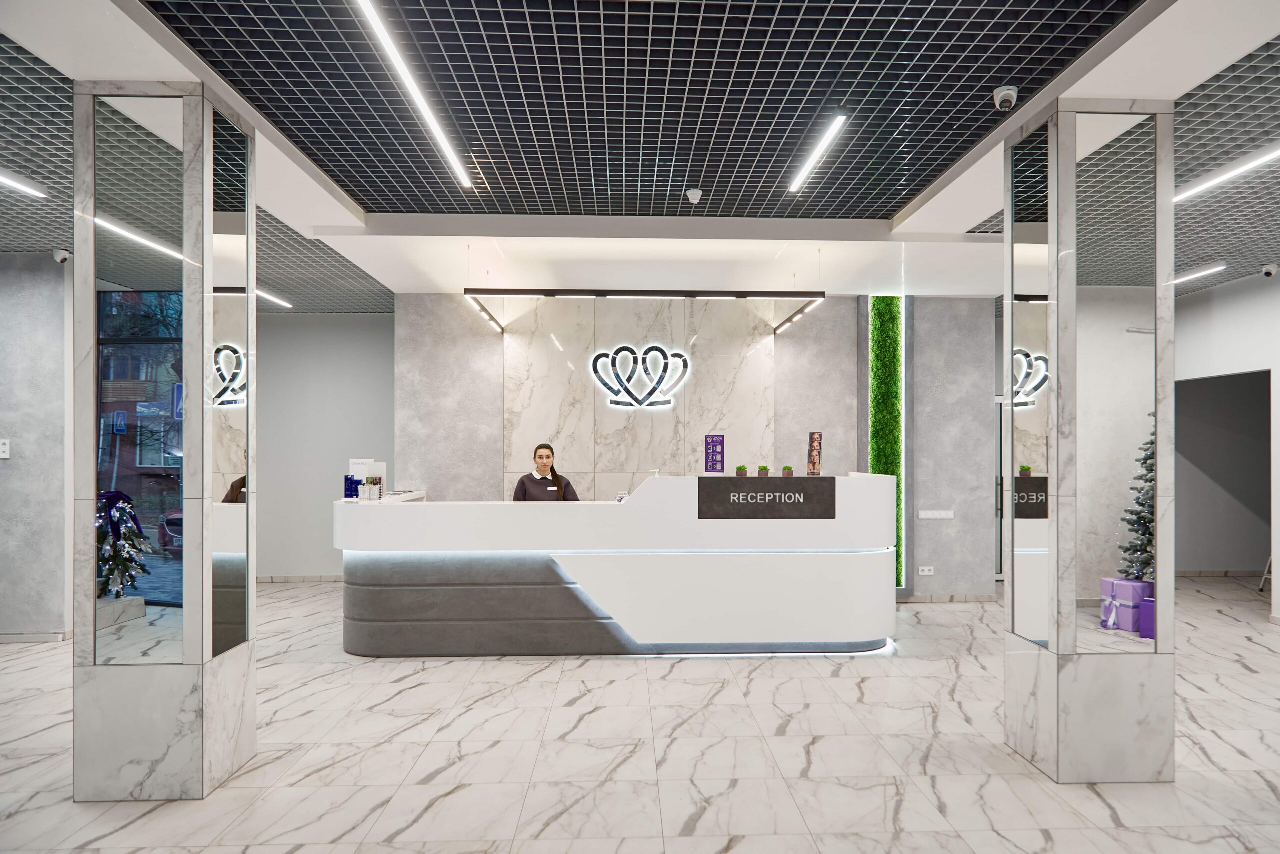
📍 Lviv, Ukraine
A dental clinic in Lviv with a separate doctor for each field: oral surgery and implants, prosthodontics, restorative dentistry, endodontics under a microscope, periodontics, orthodontics and paediatric dentistry.
A panoramic X-ray produces a single flat image of both jaws: the position of the teeth, the height of the bone, the general picture. CT builds a three-dimensional volume and shows slices in three planes, including the width of the bone from cheek to tongue, which a panoramic image cannot show at all. CT involves a higher dose, so it is prescribed when three-dimensional information is genuinely needed: before implant placement, for a complex extraction, or when close contact between a root and the mandibular canal is suspected.
At the clinics MediUkraine works with, a panoramic X-ray of both jaws costs from USD 9 including the image file. A CT scan of both jaws is charged separately, from USD 15 a dentist consultation from USD 8 and a written treatment plan from USD 14. Some clinics charge separately for a detailed radiologist's report. The clinic confirms the final cost.
The dose depends on the unit and the imaging mode and is usually measured in single to tens of microsieverts, which is comparable to a few days of natural background radiation. Digital units need a lower dose than film-based ones. That does not mean X-rays can be taken "just in case": every examination should answer a specific question from the dentist, and if you have a recent image from another clinic, it is simpler to bring it than to take a new one.
Children can, usually from the age of five or six, once they can stand still for 15–20 seconds; units have a paediatric programme with a reduced field. In mixed dentition this image is often essential: it is used to count the permanent tooth buds and check that they are all present. During pregnancy, routine X-rays are postponed; if the examination is needed because of an acute problem, the decision is made together with the patient: the beam is aimed at the jaws, and the abdomen is outside the field.
Usually not. A two-dimensional image shows the height of the bone and the distance between neighbouring roots, but not the width of the ridge from cheek to tongue, the concavity on the inner side of the lower jaw, the septa in the sinus or the exact course of the mandibular canal. Combined with variable horizontal magnification, this makes measurements unreliable. That is why cone beam CT is done before implant placement, sinus lifts and bone grafting, while the panoramic X-ray remains the first step.
No special preparation is needed. Before the X-ray you remove earrings, piercings, glasses, hairpins, chains and removable dentures: metal produces a white spot and a blurred ghost shadow on the opposite side of the image. In the unit you have to stand still for 15–20 seconds, biting on a plastic block with your lips closed and your tongue pressed against the roof of your mouth; otherwise a dark band will fall across the roots of the upper teeth. The whole visit takes 5–10 minutes.
Large cavities, yes; early caries between the back teeth, mostly not. The contact surfaces of neighbouring teeth overlap one another, and the resolution of a panoramic X-ray is lower than that of periapical X-rays. To look for caries at the contact points, bitewings are taken: images of the crowns of the upper and lower back teeth in a single frame. So a panoramic image complements a dental examination and periapical X-rays rather than replacing them.
Tell us what you need: we will suggest a clinic and a doctor and send you a cost estimate.
Request a consultation →