
| Service | 🇺🇦 Ukraine | 🇨🇿 Czech Republic | 🇹🇷 Turkey |
|---|---|---|---|
| Tooth extraction | from USD 42 | from USD 73 | from USD 52 |
| Wisdom tooth extraction | from USD 52 | from USD 143 | from USD 167 |
| Computer-controlled anaesthesia | from USD 13 | from USD 7 | from USD 7 |
| Emergency pain relief | from USD 24 | from USD 72 | from USD 35 |
Prices in the “Ukraine” column come from MediUkraine’s partner clinics. The Czech Republic and Turkey columns show the lower bounds of prices published by clinics there (for Turkey, also the Turkish Dental Association’s recommended 2026 tariff), collected in August 2026 and converted to US dollars at the rate on the day of collection. A dash means we could not find a comparable published price. In any country, the final cost is set by the clinic after an examination.
Tooth extraction is the surgical removal of a tooth together with its root from the bony socket. Dentists regard it as a last resort: a natural root maintains bone volume and spreads chewing forces, and no restoration fully reproduces this. So the right order is: first the dentist assesses the chances of saving the tooth on a periapical X-ray or a cone beam CT scan, and only then discusses surgery.
A tooth can no longer be saved in these cases: a longitudinal crack or fracture of the root, destruction of the crown below the bone level with no possibility of surgical crown lengthening, grade three mobility due to periodontitis when the support has been lost, blocked or perforated canals with a focus of inflammation after unsuccessful repeat treatment, a tooth in the line of a jaw fracture. Orthodontic extraction is a separate case: premolars are removed to make room for straightening the teeth.
Teeth can be saved more often than it seems when you are in acute pain. Repeat endodontic treatment gives a predictable result even after an unsuccessful first attempt; with a cyst at the root tip, an apicectomy is performed; with deep destruction, surgical crown lengthening or orthodontic extrusion of the root is used; with mobility due to periodontitis, the teeth are splinted. These options take longer and sometimes cost more than an extraction; the choice is made with you after discussing the prognosis.
A planned extraction is postponed, or agreed with the relevant specialist, during an acute infection, within the first six months after a heart attack or stroke, in uncontrolled diabetes or acute blood disorders, and during radiotherapy to the head and neck. Taking anticoagulants, bisphosphonates or denosumab does not rule out surgery but requires special preparation, and you must not stop these medicines on your own. For pregnant women, planned procedures are usually moved to the second trimester, while emergency care is provided at any stage of pregnancy.
Wisdom teeth have their own indications and their own surgical technique, and are covered on a separate page. MediUkraine is a treatment search and coordination service, not a clinic: we match you with a clinic and an oral surgeon, arrange the date and support you throughout, including if you are travelling from abroad. The decision on whether to extract or treat the tooth is made by the dentist after an in-person examination.

Travel. Ukrainian airspace is closed, so there are no commercial flights. Patients reach Lviv overland, most often from Poland: Rzeszów and Kraków airports are the usual entry points, with a train, bus or private transfer onward to the border and on to the city. Lviv is roughly 70 km from the Polish border, the shortest crossing of any Ukrainian city.
Visas. Citizens of the EU, the United Kingdom and the United States do not need a visa for short stays in Ukraine. Rules depend on your citizenship, so check your own before booking.
Safety, plainly. Ukraine is at war. Air raid alerts occur in Lviv, and an appointment can be interrupted by one. Several governments advise against travel to Ukraine, and while that advice stands, ordinary travel and medical insurance is often void — confirm your cover with your insurer before you commit to anything.
Language. MediUkraine handles correspondence, scheduling, the written treatment plan and the quote in English. The clinic's team communicates with international patients in English.
Payment. You pay the clinic directly. Our coordination service is free of charge for you.
More on travel, safety and insurance: Safety and travel.
There are two basic types of extraction. A simple extraction is performed with forceps and elevators when the crown is intact, the roots are straight and the surrounding bone is unchanged: the surgeon cuts the circular ligament, luxates the tooth and removes it in one piece. A complex extraction requires an incision in the gum, raising a flap, partial removal of bone and sectioning the crown into fragments; this approach is used for multi-rooted molars, teeth broken down to the gum line and roots left under the gum.
The type of anaesthesia depends on the area. In the upper jaw an infiltration is usually enough; for lower molars an inferior alveolar nerve block is needed. The anaesthetic is most often articaine, with epinephrine at a dilution of 1:100,000 or 1:200,000; for patients with cardiovascular disease a lower concentration of the vasoconstrictor or a solution without it is chosen. Computer-controlled anaesthesia delivers the solution slowly and under pressure control, so the injection is felt less, which makes it a sensible choice if you are afraid of injections. Sedation relieves anxiety but does not replace local anaesthesia.
The atraumatic technique means the surgeon works by separating rather than rocking the tooth. The periodontal ligament is cut around the root with a periotome or luxator, gradually going deeper, and the tooth comes out of the socket without pressure on its walls. Before this, a multi-rooted molar is sectioned into separate roots, each removed in turn, which preserves the thin buccal wall and the inter-radicular septum on which the future bone volume depends.
Piezosurgery uses an ultrasonic tip that cuts mineralised tissue without damaging soft tissue. It is used where a root lies against the mandibular canal, the floor of the maxillary sinus or a neighbouring implant. The method is slower than a bur, so it is not used every time, only when the anatomy calls for it.
Once the root is out, the socket is always cleaned and checked. Granulation tissue, remnants of the periodontal ligament and the cyst lining, if there was one, are removed with a surgical curette; sharp bone edges are smoothed and the wound is rinsed with an antiseptic solution. Granulation tissue left behind is one of the most common reasons a socket stays painful and heals poorly.
Socket preservation makes sense when an implant is planned in place of the tooth. After the root is lost, the bone remodels: in the first few months the ridge loses noticeable width, and the thin outer wall suffers most. To compensate for this loss, the socket is filled with bone graft material (a xenograft from bovine bone, an allograft or a synthetic tricalcium phosphate material) and covered with a collagen membrane or sponge. Another option is a membrane prepared from the patient's own blood by centrifugation.
Immediate implant placement means placing an implant in the same socket straight after the root has been removed. It is possible when there is no acute purulent inflammation, the socket walls are intact, and there is enough of the patient's own bone beyond the root tip for primary stability. In the front of the mouth this approach helps preserve the gum contour, but it does not suit everyone: if the buccal wall is destroyed or there is active infection with pus, the socket is allowed to heal first and the implant is placed later.
A gap in the smile line is filled with a temporary restoration. The simplest is a removable immediate denture, which is made in advance and fitted straight after the operation; it is for appearance, not for chewing. Further back in the mouth a temporary replacement is usually unnecessary if the permanent restoration is planned within a few months.
The edges of the wound are brought together with sutures. Absorbable sutures dissolve on their own within one to two weeks; non-absorbable ones are removed on day 7–10, which is a quick and almost painless procedure. Not every socket needs sutures: after a simple extraction of a single-rooted tooth a sterile swab and a few minutes of biting down are often enough.
Impacted and malpositioned wisdom teeth require a different protocol and separate planning based on a CT scan, and are covered on a separate page. Conversely, if it turns out at the consultation that the tooth can still be saved by repeat root canal treatment, an apicectomy or splinting, the surgeon will say so directly. Only a dentist who has examined you and reviewed the X-ray can predict the outcome in your case.
The first 24 hours determine how the socket will heal. Bite on the sterile swab for 20–30 minutes, then remove it; there is no need to keep it longer, as blood-soaked gauze itself becomes a breeding ground for bacteria. A blood clot forms in the socket, and it is this clot that protects the bone: on the first day you must not rinse your mouth, drink through a straw, apply heat to the cheek or smoke. For the first two to three hours, hold something cold against the cheek for 10 minutes at a time, with breaks of the same length.
Pain after the procedure is expected and is usually controlled with ordinary painkillers, which the dentist will recommend at the appointment. Swelling builds until the second day, stays on the third and then goes down; sometimes a bruise appears on the skin, which fades within a week and a half. A temperature of up to 37.5 °C on the first day is a normal reaction to surgery, but a temperature that persists into the third or fourth day needs checking by a dentist.
You can eat after 2–3 hours, once the anaesthetic has worn off: food should be soft and warm rather than hot, and it is best to chew on the opposite side. Brush your teeth from the next morning with a soft brush, avoiding the extraction site. From the second day, use mouth baths rather than rinses: take the antiseptic solution into your mouth, hold it for half a minute and gently let it out without creating suction.
Healing times differ for the gum and for the bone. The gum over the socket closes in 7–10 days, which is when the sutures are removed; young bone fills the socket in about 3–4 months, and its full remodelling takes up to six months. Everything looks healed from the outside much sooner than the process is complete inside, which is why the timing of the restoration is based on an X-ray, not on how the gum looks.
If an implant is planned, the timing depends on the clinical situation. Early delayed implantation is done after 6–8 weeks, when the gum has healed but the bone is still actively remodelling; the classic delayed option is after 3–4 months. After the socket has been filled with bone graft material, the wait is longer, usually 4–6 months, and the CT scan is repeated before the operation.
See a dentist without waiting for your next appointment if the pain is not easing but increasing from the second or third day and radiates to the ear or temple, if a foul taste appears, if bleeding does not stop for more than two hours, or if swelling makes it hard to open your mouth and swallow. A routine check-up is scheduled for day 2–3 and for the day the sutures are removed. If needed, MediUkraine puts you in touch with the surgeon after you return home.
Step 1. Examination and X-ray. The surgeon examines the tooth and surrounding tissues, assesses mobility and the condition of the gums, and studies the periapical X-ray or CT scan: how many roots there are, whether they are curved, how thick the outer wall of the socket is, how close the mandibular canal and the floor of the maxillary sinus lie. At this stage the final decision is made on whether the tooth can be saved and whether socket preservation is needed for a future implant.
Step 2. Anaesthesia. The gum is treated with a topical gel, then the anaesthetic is injected, by infiltration in the upper jaw or as a nerve block in the lower. After 5–10 minutes the dentist checks the depth of anaesthesia and starts only when you can no longer feel the instrument. If needed, more anaesthetic is given during the procedure.
The whole visit takes about 40–60 minutes, of which the operation itself accounts for 20–40. Between the anaesthetic and suturing, the surgeon separates the periodontal ligament, removes the root whole or in parts, cleans the socket of granulation tissue and, if needed, fills it with bone graft material for a future implant. There is no need to stay in hospital: you go home the same day with a swab, aftercare instructions and a follow-up date.
The cycle begins with a diagnostic visit. The surgeon looks at the tooth and the X-ray, involves a general dentist or prosthodontist if needed, and draws up a plan: save the tooth, operate now, or first relieve the flare-up. If the situation is simple and there are no contraindications, the procedure is done the same day.
The second stage is the operation itself and the two days that follow. During this time it is important not to miss the follow-up examination on day 2–3: the dentist checks whether a clot has formed and whether there are signs of inflammation, and adjusts the prescriptions if needed.
The third stage is suture removal on day 7–10 and a check on how the gum has healed. At this visit the options for replacing the tooth are discussed, because it is now clear how the socket has healed and how much bone has been preserved.
Next comes the period of bone remodelling, from several weeks to several months depending on whether the socket was filled with material. At the end of this period a follow-up CT scan is taken and an implant or a bridge is planned. If you decide not to replace the tooth, the dentist explains the consequences: the neighbouring teeth gradually tilt towards the gap, and the opposing tooth over-erupts towards it.
If you are travelling from abroad, it is convenient to split treatment into two trips: the first for diagnostics, the operation, a follow-up and suture removal, which takes about 7–10 days; the second for implantation or prosthetic treatment a few months later. MediUkraine keeps your treatment records between trips and times the second visit for when the bone is ready.
The most common complication is dry socket (alveolitis), inflammation of the socket walls after the blood clot is lost. It develops on the second to fourth day: the pain does not subside but intensifies and radiates to the ear, temple or neck, a foul smell appears, and the socket looks empty or covered with a grey coating. The risk is increased by smoking, rinsing on the first day, drinking through a straw, traumatic surgery and inadequate hygiene. Dry socket is treated at the clinic: the socket is rinsed, necrotic tissue is removed and a medicated dressing is placed, after which the pain usually subsides within one to two days. Self-treatment with painkillers only delays the visit.
Bleeding from the socket in the first hours is expected, but if it does not stop for more than two hours an examination is needed; sometimes a suture or a haemostatic sponge has to be placed. Patients on anticoagulants are a separate case: their bleeding lasts longer, and the approach is agreed in advance with the doctor who prescribed the medicine. Limited mouth opening and a bruise on the cheek resolve on their own within one to two weeks.
When lower molars are removed there is a risk of injury to the inferior alveolar or lingual nerve, with numbness of the lip, chin or half of the tongue. Most often this is the result of compression and swelling rather than a tear, so sensation returns within a few weeks or months; persistent problems are rare. This is why a CT scan is taken before a complex extraction in the lower jaw: to assess the risk in advance.
The roots of the upper molars and premolars are sometimes separated from the floor of the maxillary sinus only by a thin plate of bone, so a sinus perforation is possible. The signs are air passing through the socket and liquid getting into the nose. The opening is closed immediately, and you must not blow your nose, fly or dive for two weeks. Less common are fracture of the socket wall, displacement of a root fragment and chipping of a restoration on a neighbouring tooth; some roots break during the procedure, and the surgeon then completes the extraction through a surgical (open) approach.
Particular attention is paid to medicines that affect bone. Bisphosphonates and denosumab, prescribed for osteoporosis and for bone involvement in cancer, raise the risk of medication-related osteonecrosis of the jaw, so before the operation a consultation with the prescribing doctor is needed, together with written consent after the risk has been discussed. The same approach applies after radiotherapy to the head and neck. In uncontrolled diabetes a planned extraction is postponed until blood glucose is under control, and during an acute infection until you have recovered.
The risk of delay is real too. A tooth with a chronic focus at the root tip remains a source of infection, and a flare-up can progress to periostitis or an abscess, when surgery has to be done urgently and in less favourable conditions. At the same time, hasty extraction of a tooth that could still have been saved is irreversible: there will be no second attempt. That is why you are entitled to a second opinion: the X-ray and the examination record can be shown to another specialist, and if needed MediUkraine arranges a second consultation at another partner clinic.
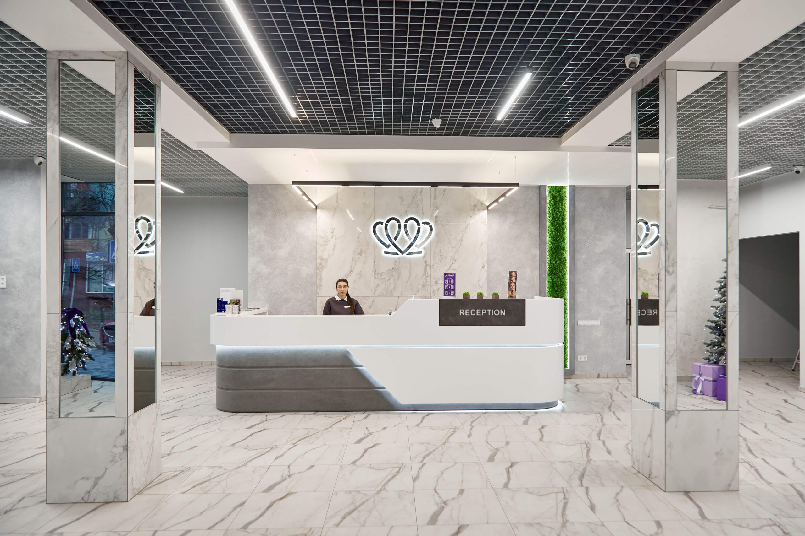
📍 Lviv, Ukraine
A dental clinic in Lviv with a separate doctor for each field: oral surgery and implants, prosthodontics, restorative dentistry, endodontics under a microscope, periodontics, orthodontics and paediatric dentistry.
There is no pain during the operation: the area is numbed by infiltration or a nerve block, and the surgeon starts work only after checking the depth of anaesthesia. You will feel pressure and movement, but not pain. If the numbness starts to wear off, say so straight away and more anaesthetic will be given. Discomfort comes later, once the anaesthetic wears off, and is usually controlled with ordinary painkillers for two to three days. For those with a marked fear of injections, computer-controlled anaesthesia or sedation is offered.
At the clinics MediUkraine works with, a simple extraction starts from USD 42 and a wisdom tooth extraction from USD 52. Computer-controlled anaesthesia is charged separately, from USD 13, and an emergency pain relief appointment from USD 24. An X-ray or CT scan, bone graft material for socket preservation and implantation are paid for as separate items. The clinic sets the final cost after an examination and imaging, so each quote is individual.
More often than it seems when you are in pain. Repeat root canal treatment helps even after an unsuccessful first attempt; with a cyst at the root tip, an apicectomy is performed; with deep destruction, the crown is lengthened surgically or the root is extruded orthodontically; with mobility due to periodontitis, the teeth are splinted. A tooth is considered hopeless with a longitudinal root crack, destruction below the bone level that cannot be restored, or grade three mobility. The decision is made by the dentist based on an X-ray or CT scan.
It depends on the condition of the socket. Sometimes the implant is placed straight into the same socket, if there is no purulent inflammation, the walls are intact and there is enough bone beyond the root tip. Early delayed implantation is done after 6–8 weeks, the classic delayed option after 3–4 months. If the socket was filled with bone graft material, the wait is longer, usually 4–6 months. The CT scan is repeated before the operation to assess the actual bone volume.
Bite on the swab for 20–30 minutes, then remove it. On the first day you must not rinse your mouth, drink through a straw, smoke, take a hot bath or exercise: all of these can dislodge the blood clot in the socket. Apply cold to the cheek for 10 minutes at a time, with breaks of the same length. You can eat after 2–3 hours: food should be soft and warm, and it is best to chew on the opposite side. Brush your teeth from the next morning, avoiding the extraction site, and from the second day use antiseptic mouth baths instead of rinses.
A dry socket is alveolitis, inflammation of the socket walls after the blood clot has been lost. Typical signs: on the second to fourth day the pain does not ease but increases, radiating to the ear or temple, an unpleasant taste and smell appear, and the socket looks empty or grey. This cannot be treated with painkillers at home: at the clinic the socket is rinsed, necrotic tissue is removed and a medicated dressing is placed, after which the pain usually subsides within a day or two. The best prevention is not to smoke or rinse your mouth on the first day.
Not always. Filling the socket with bone graft material is done primarily when an implant is planned at that site and preserving the width of the ridge matters. If the gap is not going to be replaced, the socket heals on its own, although the bone loses some of its volume over time. It is worth understanding the consequences of not replacing the tooth: the neighbouring teeth gradually tilt towards the gap, the opposing tooth over-erupts towards it, and the load on the rest of the arch increases. The dentist will tell you what makes sense in your case after an examination.
Tell us what you need: we will suggest a clinic and a doctor and send you a cost estimate.
Request a consultation →