| Service | 🇺🇦 Ukraine | 🇨🇿 Czech Republic | 🇹🇷 Turkey |
|---|---|---|---|
| Bone grafting | from USD 144 | from USD 239 | from USD 334 |
| Closed sinus lift | from USD 489 | from USD 239 | from USD 290 |
| Open sinus lift | from USD 733 | from USD 716 | from USD 580 |
| CT scan (2 jaws, for clinic patients) | from USD 15 | — | — |
Prices in the “Ukraine” column come from MediUkraine’s partner clinics. The Czech Republic and Turkey columns show the lower bounds of prices published by clinics there (for Turkey, also the Turkish Dental Association’s recommended 2026 tariff), collected in August 2026 and converted to US dollars at the rate on the day of collection. A dash means we could not find a comparable published price. In any country, the final cost is set by the clinic after an examination.
Jawbone needs load to survive. While a tooth is in place, chewing forces pass through the root into the alveolar bone and maintain its volume; once the tooth is extracted, that load disappears and the bone starts to shrink. Most of the loss happens in the first months: within a year the ridge loses between a quarter and a half of its original width, after which the process slows down but does not stop.
An implant needs a certain amount of bone. At least 1 mm of natural bone must surround it on each side, with a safe distance from its tip to the mandibular canal and the floor of the maxillary sinus. In practice, a standard-diameter implant needs a ridge about 6 mm wide or more; when bone loss leaves only 3–4 mm, there is no room for an implant, and that is when grafting is needed.
Indications include teeth that have been missing for a long time, a traumatic extraction that fractured the socket wall, the after-effects of cysts, granulomas and chronic periodontitis, bone loss from gum disease, jaw injuries and congenital defects. The back of the upper jaw is a special case: the sinus expands downwards over time, leaving 2–5 mm of bone height, and a sinus lift is used there (it has its own page in our catalogue).
The procedure is not suitable for everyone. Planned grafting is postponed or ruled out in the case of untreated gum disease and poor hygiene, uncontrolled diabetes, acute inflammation in the mouth, pregnancy, during chemotherapy, after radiotherapy to the head and neck, and in patients receiving intravenous bisphosphonates or denosumab injections, in whom medication-related osteonecrosis of the jaw is possible. Heavy smoking does not formally rule out the procedure, but it noticeably worsens the outlook, and the surgeon will discuss this with you beforehand.
MediUkraine is a treatment search and coordination service, not a clinic. We choose the clinic and a surgeon with the right specialism, arrange the dates and support you throughout, including if you are travelling from abroad. The scope of the procedure, the technique and the material are decided by the doctor after an in-person examination and a review of the CT scan.

Travel. Ukrainian airspace is closed, so there are no commercial flights. Patients reach Lviv overland, most often from Poland: Rzeszów and Kraków airports are the usual entry points, with a train, bus or private transfer onward to the border and on to the city. Lviv is roughly 70 km from the Polish border, the shortest crossing of any Ukrainian city.
Visas. Citizens of the EU, the United Kingdom and the United States do not need a visa for short stays in Ukraine. Rules depend on your citizenship, so check your own before booking.
Safety, plainly. Ukraine is at war. Air raid alerts occur in Lviv, and an appointment can be interrupted by one. Several governments advise against travel to Ukraine, and while that advice stands, ordinary travel and medical insurance is often void — confirm your cover with your insurer before you commit to anything.
Language. MediUkraine handles correspondence, scheduling, the written treatment plan and the quote in English. The clinic's team communicates with international patients in English.
Payment. You pay the clinic directly. Our coordination service is free of charge for you.
More on travel, safety and insurance: Safety and travel.
Guided bone regeneration is the core technique: bone material is placed in the deficient area and covered with a barrier membrane. The membrane is there not for strength but to keep tissues apart. Gum epithelium and connective tissue grow several times faster than bone and, without a barrier, would simply fill the space meant for new bone; under the membrane, the clot matures into bone undisturbed over months. That is why an intact membrane and watertight stitches matter as much as the material itself.
Autogenous bone, your own bone, is the only material containing living bone-forming cells. It is taken near the surgical site: from the external oblique ridge or ramus of the lower jaw, the retromolar area or, less often, the chin; small amounts are collected with a bone scraper or a filter trap during drilling. Autogenous bone matures fastest but has two limitations: only a small volume is available, and it resorbs noticeably, so it is usually mixed roughly half and half with a slow-resorbing material rather than used alone.
Xenogeneic granules are bovine or porcine bone with all proteins removed, leaving a naturally porous mineral framework. This material barely resorbs and maintains the gained volume for years, so it is used when the ridge shape needs to be preserved long term. Allogeneic materials are processed human donor bone from a tissue bank. Alloplasts based on beta-tricalcium phosphate and hydroxyapatite are fully synthetic: the body replaces them with its own tissue, but they also lose volume faster.
Membranes are divided into resorbable and non-resorbable. A resorbable collagen membrane dissolves over several weeks or months and does not need removing; it is used for small and medium defects. Non-resorbable PTFE membranes, often titanium-reinforced, and titanium meshes hold their shape even under pressure from the soft tissues, so they are essential for large reconstructions; they are fixed with pins or microscrews and removed in a separate procedure several months later. The trade-off for rigidity is a higher risk of the membrane edge becoming exposed through the stitches.
Alveolar ridge splitting is used when bone height is sufficient but the ridge is narrow, about 3–5 mm. The ridge is cut lengthwise with a piezo scalpel or thin discs, the outer and inner walls are gently spread apart, and the gap between them is filled with graft material. The method gains width immediately, often allows an implant to be placed at the same time and shortens treatment by months. It needs a layer of spongy bone between the cortical plates, so it is not always possible on a thin, knife-edge lower ridge.
When the defect is large, a bone block is used: a piece of your own bone from the ramus of the lower jaw, or an allogeneic block, is fitted against the ridge and fixed with microscrews, and the gaps are filled with granules and covered with a membrane. A block gives predictable width but is more invasive because of the second surgical site. The most challenging area is vertical augmentation, when the ridge lacks height: titanium mesh, tenting screws and sometimes distraction osteogenesis are used, and surgeons openly acknowledge that vertical gain is less predictable than horizontal gain and more often comes with membrane exposure.
Socket preservation is worth knowing about separately. If bone material is placed in the socket straight after an extraction and covered with a membrane, the ridge loses much less volume than with normal healing. It is a short procedure done at the same visit, and it often avoids the need for full grafting a year later. If you are having a tooth out and plan an implant later, ask about this option before the extraction.
The key planning decision is whether to graft separately or at the same time as placing the implant. If the deficit is small and the implant can go in the correct position with enough primary stability, both are done in one visit. If there is so little bone that the implant will not hold or will sit at the wrong angle, the stages are separated: bone first, then the implant once the graft has matured. Trying to save a visit in this situation usually ends in losing the implant.
Healing times depend on the size and type of the defect. Small horizontal defects and sockets are ready after about 4 months, large horizontal ones after 6, and vertical reconstructions and blocks after 6–9 months. Readiness is judged not by the calendar but by a follow-up CT scan: the doctor checks whether density and width are sufficient for an implant of the required size. Platelet-rich fibrin and membranes made from your own blood help the soft tissues heal, but on their own they do not add volume.
There is no one-size-fits-all method, and it is worth knowing this before you pay. The choice between ridge splitting, granules under a membrane and a block depends on the shape of the defect, gum thickness, the condition of the neighbouring teeth, your habits and the planned restoration; the same millimetre deficit is treated differently in the upper and lower jaw. Only a doctor, after an examination and a review of the CT scan, can predict the outcome in your case, not remotely from a description or a smile photo.
Expect swelling for the first two to three days. It builds for 48–72 hours, then subsides, and by day 5–7 the face usually looks normal again; a bruise on the cheek or under the jaw may appear and fades on its own. Apply cold only on the first day, 15 minutes at a time with breaks; after that it no longer helps.
Take painkillers on the schedule the surgeon gives you, and finish the full course of any antibiotic prescribed. Do not rinse until the next day, so as not to dislodge the clot; after that you will be prescribed gentle chlorhexidine mouth baths, and the stitched area is cleaned carefully with a soft brush so that no plaque builds up. Avoid saunas, hot baths, the gym and straining or bending over for the first week.
After surgery on the upper jaw, do not blow your nose hard, puff out your cheeks, dive or play wind instruments for about two weeks: a change in pressure can shift the graft material. Sneeze with your mouth open. These restrictions are stricter if a sinus lift was done at the same time.
For the first two weeks, eat soft food that is not hot, and do not chew on the treated side. Stitches come out on day 7–14, sometimes later if a non-resorbable membrane was used. Do not wear a removable denture over the treated area until the doctor says so, or have it relined with a soft material so that it does not press on the stitches.
The result is judged not by how you feel but by a follow-up CT scan after 4–6 months, or 6–9 for large defects. There is one criterion: whether there is enough width, height and density to place an implant of the required diameter in the correct position. Some of the graft resorbs in this time; this is expected and allowed for, which is why the surgeon deliberately overbuilds the volume slightly.
To be honest about the method's limits: sometimes the gain is smaller than planned, and then either a smaller implant is placed or a smaller repeat graft is done. This happens mainly with vertical reconstructions and in smokers. It is a limitation of the technique, not necessarily the surgeon's mistake, which is why timelines and likely scenarios are discussed before surgery, not after the follow-up scan.
Step 1. Examination, CT scan and plan. The surgeon compares the clinical findings with the CT scan, measures bone height and width at the future implant site and decides how much volume is missing and which method to use. The material and type of membrane are chosen at this point, along with whether an implant can be placed at the same time, and the plan is put in writing together with the quote.
Step 2. Anaesthesia and access. The procedure is done under local anaesthesia, with sedation on request. Once the area is numb, the surgeon makes an incision along the ridge and lifts a gum flap, exposing only as much bone as needed, and assesses the defect directly rather than just on the scan.
In total the procedure takes 40–90 minutes: after anaesthesia and the incision, the surgeon prepares the site, splits a narrow ridge if needed, packs in the bone material, covers it with a membrane and closes the wound without tension. You spend about two to three hours at the clinic including preparation and go home the same day; stitches come out on day 7–14, and a follow-up CT scan after 4–9 months is used to plan implant placement.
Bone grafting is not a stand-alone treatment but the first stage of a pathway that ends with a crown on an implant. It takes about 9–18 months from the first consultation to the permanent restoration, and most of that time is not the doctor's work but biology: bone heals at its own pace and cannot be hurried.
The first phase is examination and preparation: consultation, CT scan, professional cleaning, treatment of decay and gum disease and, if needed, a letter from your GP about chronic conditions and medicines. It takes from a few days to three weeks depending on the state of your mouth, and it is done before surgery, not alongside it.
The second phase is the surgery itself and soft-tissue healing: the procedure, a check on day 2–3 and stitch removal on day 7–14. Then comes the longest period, 4 to 9 months while the graft matures, during which you live normally and come in only for check-ups.
The third phase begins with a follow-up CT scan. If there is enough volume, the implant is placed and needs another 3–6 months to integrate; then a healing abutment is fitted for two to three weeks, impressions or scans are taken and the crown is made. In favourable conditions the implant is placed at the same time as the graft, which shortens the whole process by several months.
If you are coming from another city or country, treatment splits conveniently into three trips: the first for the examination, scan and surgery, timed to include stitch removal; the second for the follow-up CT scan and implant placement; the third for the crown. MediUkraine plans the schedule so that each trip is used efficiently and stays in touch between stages, including with a doctor where you live if some follow-up is easier to do at home.
The most common complication is the stitches opening and exposing the membrane in the first two weeks. The larger the graft, the more likely this is, especially with non-resorbable membranes and titanium meshes. The exposed area needs careful management, and sometimes the membrane is removed early, in which case the gain is smaller than planned.
Infection, prolonged bleeding and haematoma are also possible, and in the upper jaw, perforation of the sinus lining. At the back of the lower jaw, the inferior alveolar nerve can be irritated or injured, causing numbness of one side of the lip and chin: temporary numbness resolves within weeks or months, and permanent numbness is rare. That is why the position of the nerve canal is measured on the CT scan before surgery rather than relying on average anatomy.
Later problems include partial resorption of the graft and loss of individual particles. The material is not rejected in the immune sense: xenogeneic granules contain no proteins, autogenous bone is your own, and allogeneic tissue is processed by the tissue bank. But if the site has a poor blood supply or becomes infected, some of the material does not integrate and gradually works its way out through the wound.
The factors that most worsen the outlook are well known and listed in every protocol: smoking, uncontrolled diabetes, untreated gum disease, poor hygiene, teeth grinding (bruxism) and a removable denture pressing on the treated area. These are not abstract warnings: they explain most failures, and they are the factors you can influence yourself.
Contraindications are divided into temporary and permanent. Temporary ones include acute infections, flare-ups of chronic illness, pregnancy and breastfeeding, chemotherapy and uncontrolled medical conditions; once these are resolved, surgery is possible. Permanent or almost permanent ones are intravenous bisphosphonates and denosumab injections, a history of radiotherapy to the head and neck, severe blood clotting disorders and systemic diseases in a decompensated stage.
Without grafting, there are still options: short or narrow implants within the existing bone, tilted implants, a bridge supported by your own teeth, or a removable restoration. Each has its own cost, limitations and outlook, and they are worth weighing before surgery, not after. You have the right to a second opinion: the CT scan, the plan and the quote stay with you, and if needed MediUkraine arranges a consultation at another partner clinic.
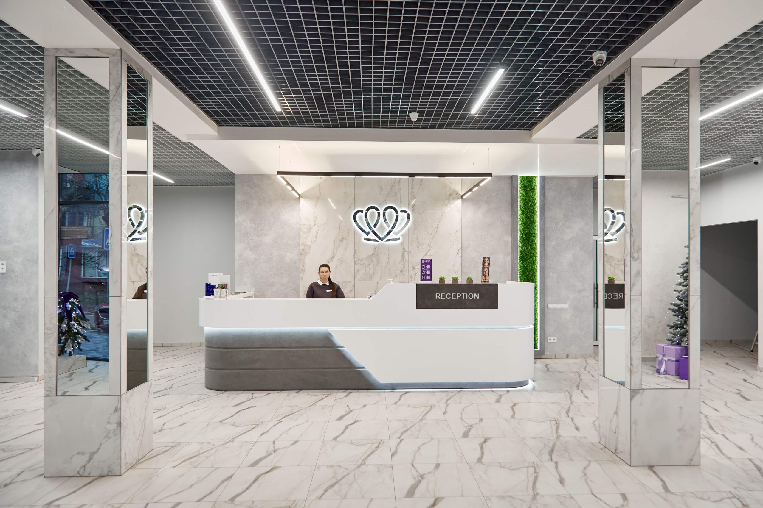
📍 Lviv, Ukraine
A dental clinic in Lviv with a separate doctor for each field: oral surgery and implants, prosthodontics, restorative dentistry, endodontics under a microscope, periodontics, orthodontics and paediatric dentistry.
When the CT scan shows the ridge is not wide or tall enough for an implant of the required size to sit in the correct position with at least a millimetre of natural bone around it. In practice this means a ridge narrower than 6 mm, too little height above the mandibular canal or below the sinus floor, or defects left by cysts, injuries and gum disease. The decision rests not on the missing tooth itself but on measurements from the scan, which is why it cannot be made remotely from a photo.
At the clinics MediUkraine works with, bone grafting costs from USD 144 per treatment area, and a cone beam CT scan of both jaws from USD 15. The bone material, membrane and fixation screws are charged separately, as the amount depends on the size of the defect. A sinus lift, if needed, is a separate item: closed from USD 489, open from USD 733. The clinic confirms the final cost after an examination and a CT scan.
It depends on the volume. Small horizontal defects and sockets preserved straight after extraction are usually ready after about 4 months; large horizontal ones after 6; vertical reconstructions and bone blocks after 6–9 months. Readiness is judged by a follow-up CT scan, not the calendar: the doctor assesses the density and true width of the ridge. When the deficit is small, the implant is placed at the same time as the graft, so there is no separate wait, just the usual 3–6 months of osseointegration.
The procedure itself is painless: it is done under local anaesthesia, with sedation if you wish. You may feel pressure and vibration, but not pain; if the anaesthetic starts to wear off, the doctor tops it up. Afterwards there may be noticeable swelling and aching for two to three days, controlled with ordinary painkillers on the prescribed schedule. Severe pain that gets worse from the third day, pus or a fever means you should call the doctor the same day rather than wait for your scheduled check-up.
Both options work, and most often they are combined. Your own bone contains living cells and matures fastest, but only a limited amount is available, harvesting it means a second surgical site, and it loses volume noticeably. Xenogeneic granules have no living cells, but they hold the shape of the ridge for years. The usual solution is therefore a mix of your own bone and a slow-resorbing material under a membrane: the first speeds up healing, the second maintains volume. The proportion and type of material are chosen by the surgeon according to the shape and size of the defect.
Yes, and this is worth knowing in advance. The material is not rejected like a transplanted organ, but it may fail to integrate because of infection, stitches opening and exposing the membrane, pressure from a removable denture, smoking or a poor blood supply. Some granules then work their way out through the wound, and the gain is smaller than planned. In that case the area is cleaned and, several months later, a smaller graft is done if needed, or a smaller implant is used instead.
There are several options, and they are chosen on the basis of the CT scan. A large deficit is treated with a bone block or vertical augmentation with titanium mesh, and at the back of the upper jaw with a sinus lift. If you are not ready for lengthy treatment, the alternatives are short or narrow implants within the existing bone, tilted implants, a bridge supported by your own teeth or a removable restoration. Each route has its own limitations in terms of lifespan and load, so they are compared before treatment begins, not after the operation.
Tell us what you need: we will suggest a clinic and a doctor and send you a cost estimate.
Request a consultation →